
Applications
What people actually do with it
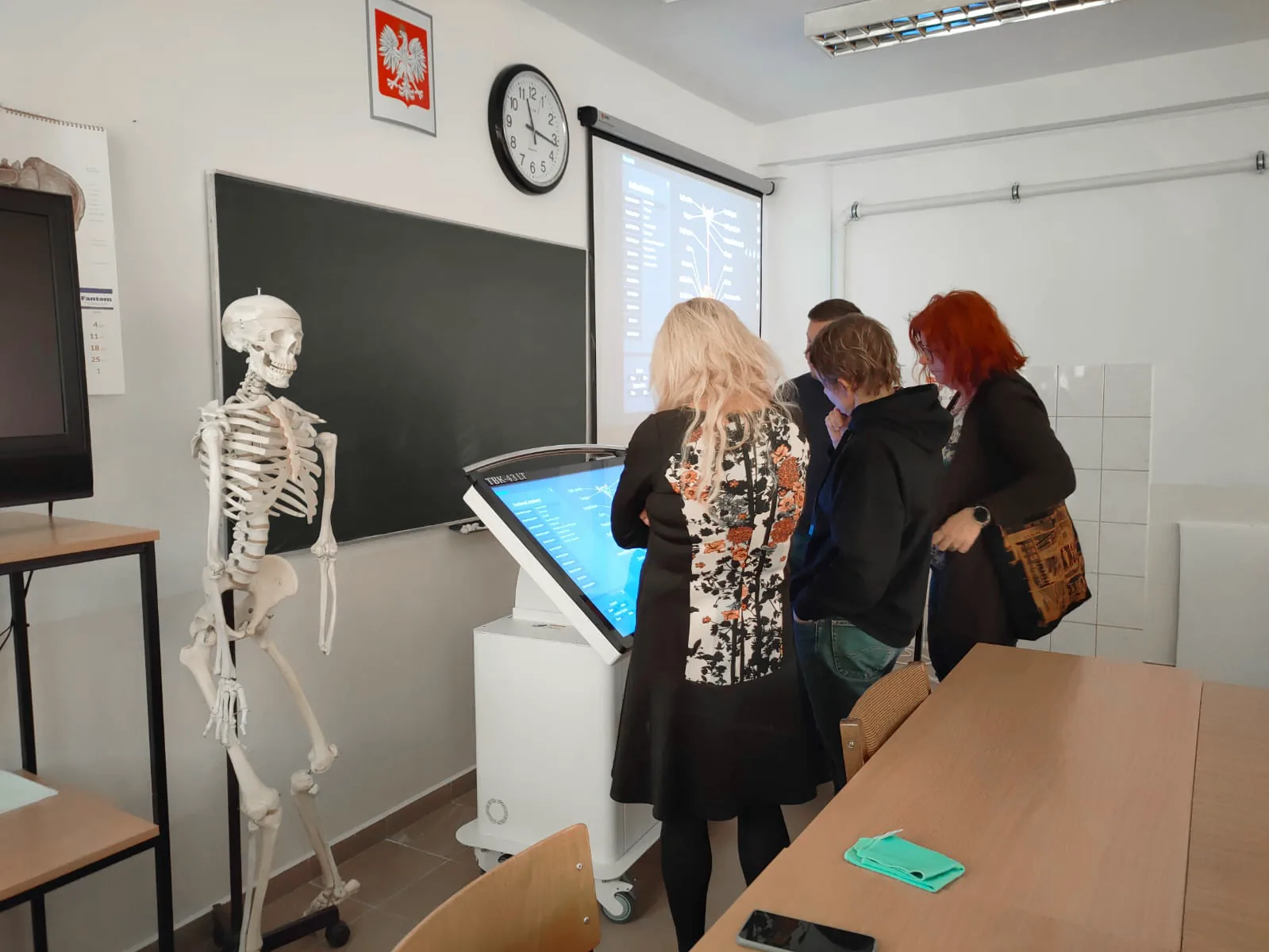
A cadaver lab that has run out of specimens. A clinician rehearsing an orthopaedic procedure on a real CT study. A consulting room where a patient finally sees what the surgeon has been describing. The Asclepius virtual dissection table is one platform put to four quite different kinds of work — this page goes through each of them, and ends with what institutions reported after they had one.
Cadaver Labs
Say goodbye to cadaver shortages
Donated specimens are scarce, expensive to keep and consumed once. A digital body is none of those things. Asclepius does not ask a department to give up dissection — it removes the ceiling on how often dissection can be practised, and takes the formalin out of the room while it is at it.
A comprehensive learning experience beyond traditional anatomy education
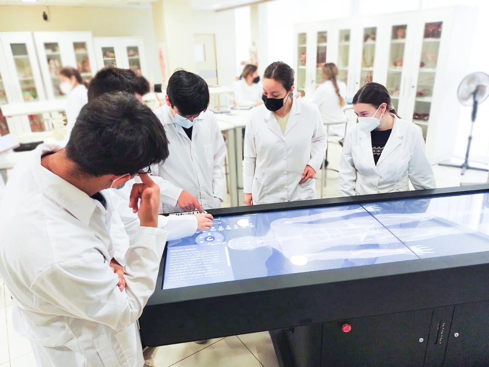
The table integrates 3D digital anatomy into the lab itself rather than parking it in a computer room next door. Students take a body apart, put it back, and take it apart again — which turns dissection from a single irreversible event into something that can be repeated until it is understood.
Nothing has to be sourced, preserved, ventilated or disposed of, so the lab is safe and clean and the number of attempts is set by the teaching, not by the supply of specimens.
Side by side with a traditional cadaver lab
| Aspect | Asclepius virtual dissection table | Traditional cadaver lab |
|---|---|---|
| Environment | No formalin odour — safe and clean | Formalin odour, exposure to harmful chemicals |
| Cadaver usage | Unlimited — digital models can be reused | Limited by physical cadaver availability |
| Cost | More cost-effective — no cadavers needed | High cost for cadavers and maintenance |
| Flexibility | Unlimited dissections on interactive 3D models | Limited by physical cadavers and dissection times |
Six things a digital body does that a preserved one cannot
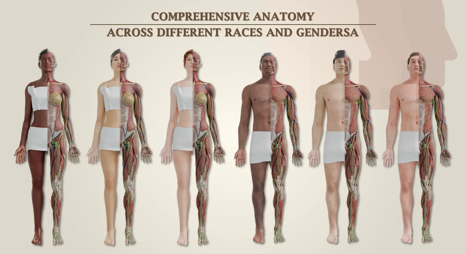
Every race, every gender
Fully digitised models covering a range of human diversity, so structures can be examined across body types instead of generalised from whichever single donor the department happens to have.
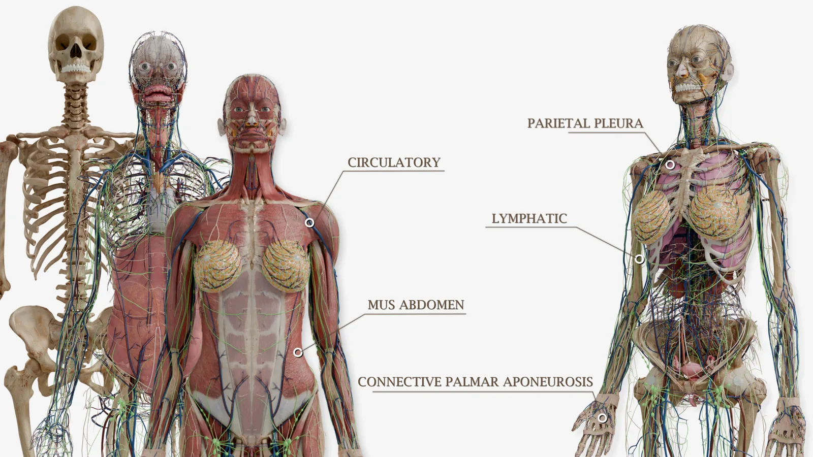
Over 5,000 anatomical structures
Every structure and physiological tissue is modelled and labelled individually, so the same body serves a gross anatomy session, a physiology lecture and a pathology seminar without anything being prepared in between.
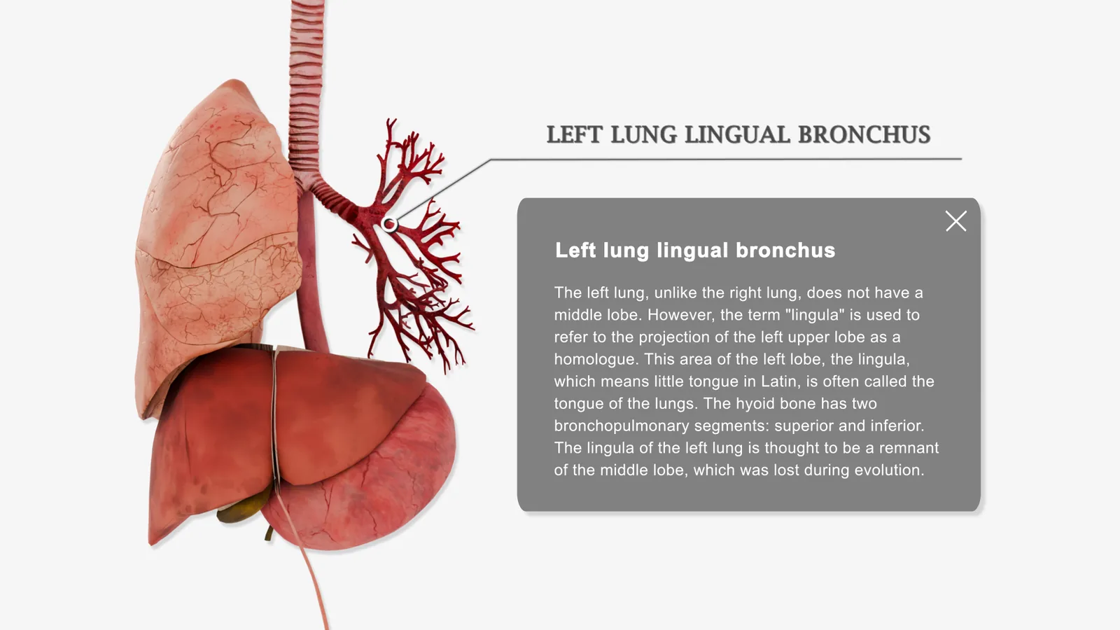
Physiology, not only anatomy
Each structure carries an explanation of what it actually does. Form and function are taught in the same breath rather than in two separate courses that the student is left to join up.
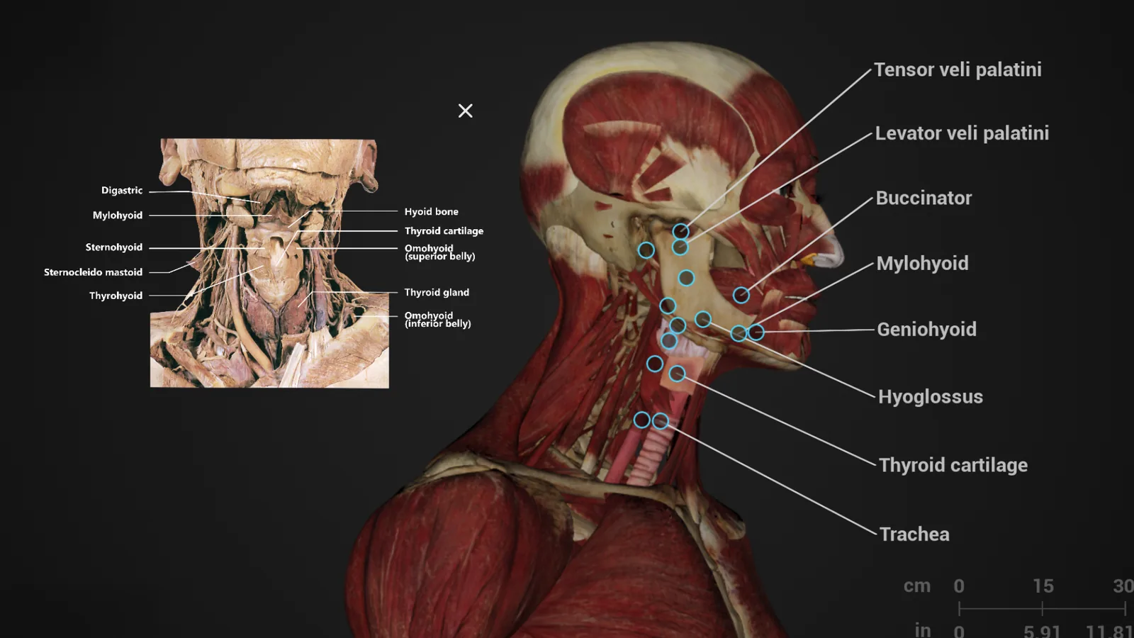
Checked against the real thing
Real anatomical photographs sit beside the digital model, so a student can validate what they have just dissected against actual human tissue rather than take the rendering on trust.
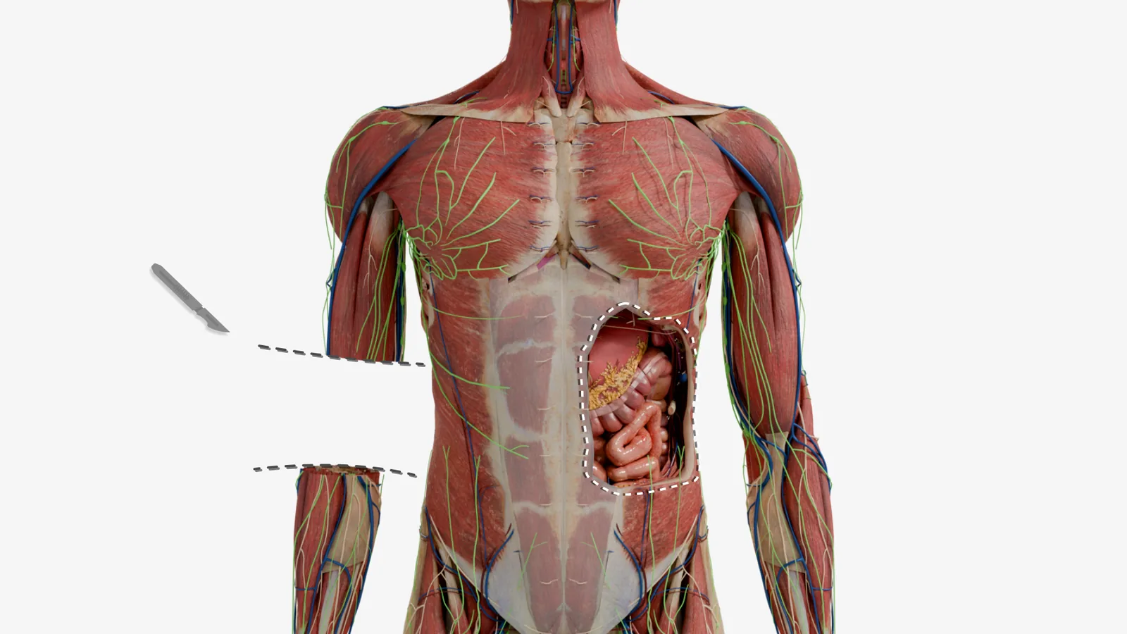
Unlimited dissections, layer by layer
Cut anywhere, peel one layer at a time, restore it and start again — a whole body or a single system. Nothing is consumed, so a difficult region can be worked through as many times as it takes.
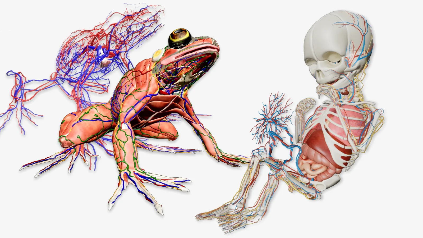
Paediatrics to veterinary anatomy
The same platform carries infant anatomy and animal specimens, so a paediatric or veterinary course does not need a second collection — or a second budget.
Whether the programme is medical, nursing or high-school biology, the argument is the same: dissection and physiology become available on demand instead of being rationed. See what each software module covers
Healthcare Professionals
Clinical practice, and the training that leads to it
For nurses, physicians and allied health staff the table is less a teaching aid than a rehearsal space. Human, embryonic and animal anatomy, normal and pathological, in 2D and 3D — and procedures that can be simulated on a virtual model before they are performed on a person.
Comprehensive anatomy education in one place
- Human, embryology and animal anatomy in a single platform, so a rounded understanding of anatomical structure does not have to be assembled from three unrelated resources
- Normal and pathological anatomy together — the complete view of human health that clinical decision-making actually requires, rather than the healthy case alone
- A 3D digital imaging platform that brings a static anatomy lesson to life: examine the body from every angle and simulate the procedure on a high-fidelity model, with no specimen involved
- Pathology and clinical diagnosis — dissect a pathological condition to understand the disease, the disorder and the treatment options, then use the same model to explain it to the patient
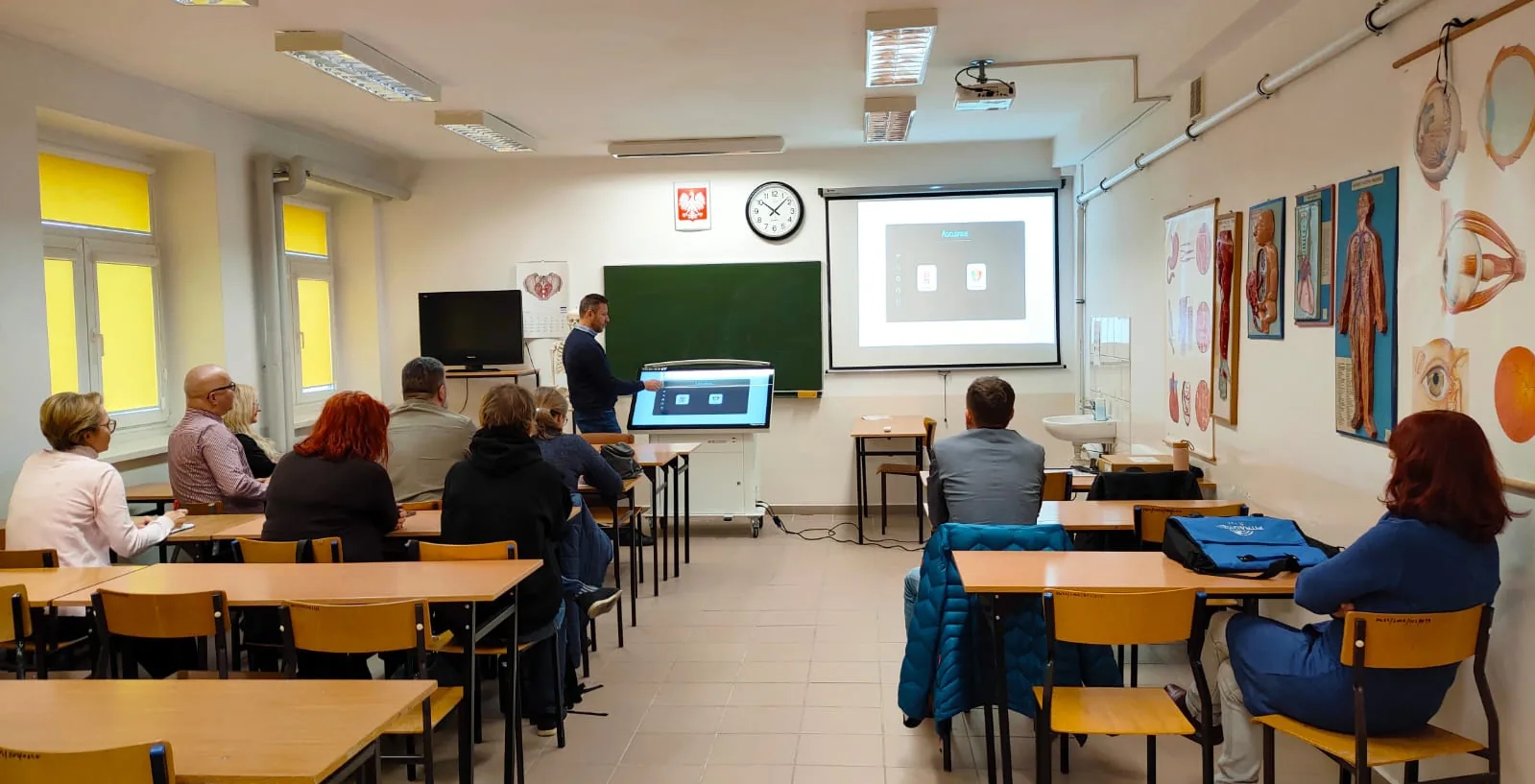
Interactive dissection and learning
Perform virtual dissections, rehearse a specific procedure and explore body systems with interactive tools — clinical skills refined without the limits of a physical cadaver.
DICOM imaging integration
Upload and visualise real DICOM studies, so training runs on the same imaging data the department already works from and diagnosis is practised at clinical accuracy.
Cost-effective and time-saving
No cadavers to buy or maintain. Training costs fall and the same resource stays available for unlimited repetition, which is where proficiency actually comes from.
In-depth learning and customisation
Select the structures or the region a session needs and leave the rest out, so practice concentrates on one body part or one system instead of the whole syllabus.
From medical imaging to surgical precision
Educators and trainees can simulate orthopaedic procedures on real clinical imaging data. High-fidelity visualisation and planning tools let students and clinicians explore, analyse and rehearse an intervention together, on the same screen.
Real CT and MRI DICOM studies replicate the clinical discussion and the preoperative plan, which turns a lecture into a case-based exercise with something genuinely at stake.
- Simulate and repeat complex procedures without physical specimens
- Reinforce anatomical knowledge through dynamic 3D visualisation
- Shorten the learning curve and improve clinical readiness
- A cost-effective alternative to running a cadaver lab
From 3D imaging to 3D printing
Rendering and physical output are one continuous path. The same study that was examined on screen can be exported as a printable model, so surgical planning runs from visualisation all the way to something the team can hold and turn over.
3D imaging files can also be uploaded and viewed in real time by several people at once — a class demonstration or a team planning session, with everyone looking at the same thing at the same moment rather than at their own copy.
See the surgical planning software
Patient Communication
Showing a patient what you are describing
Treatment goes better when the person receiving it understands it. Asclepius gives the clinician high-precision 3D imaging to explain a condition with — not a generic diagram, but the patient's own anatomy, on a screen they can see and touch.
Improved understanding, and the confidence that follows it
With the detail of the condition and the steps of the treatment laid out visually, a patient can see exactly what is happening inside their body and how it affects their health. Consent stops being a form to sign and becomes a conversation.
Patients who can see what is wrong are more comfortable with the plan for putting it right, and more likely to arrive at the procedure at ease with what has been agreed.
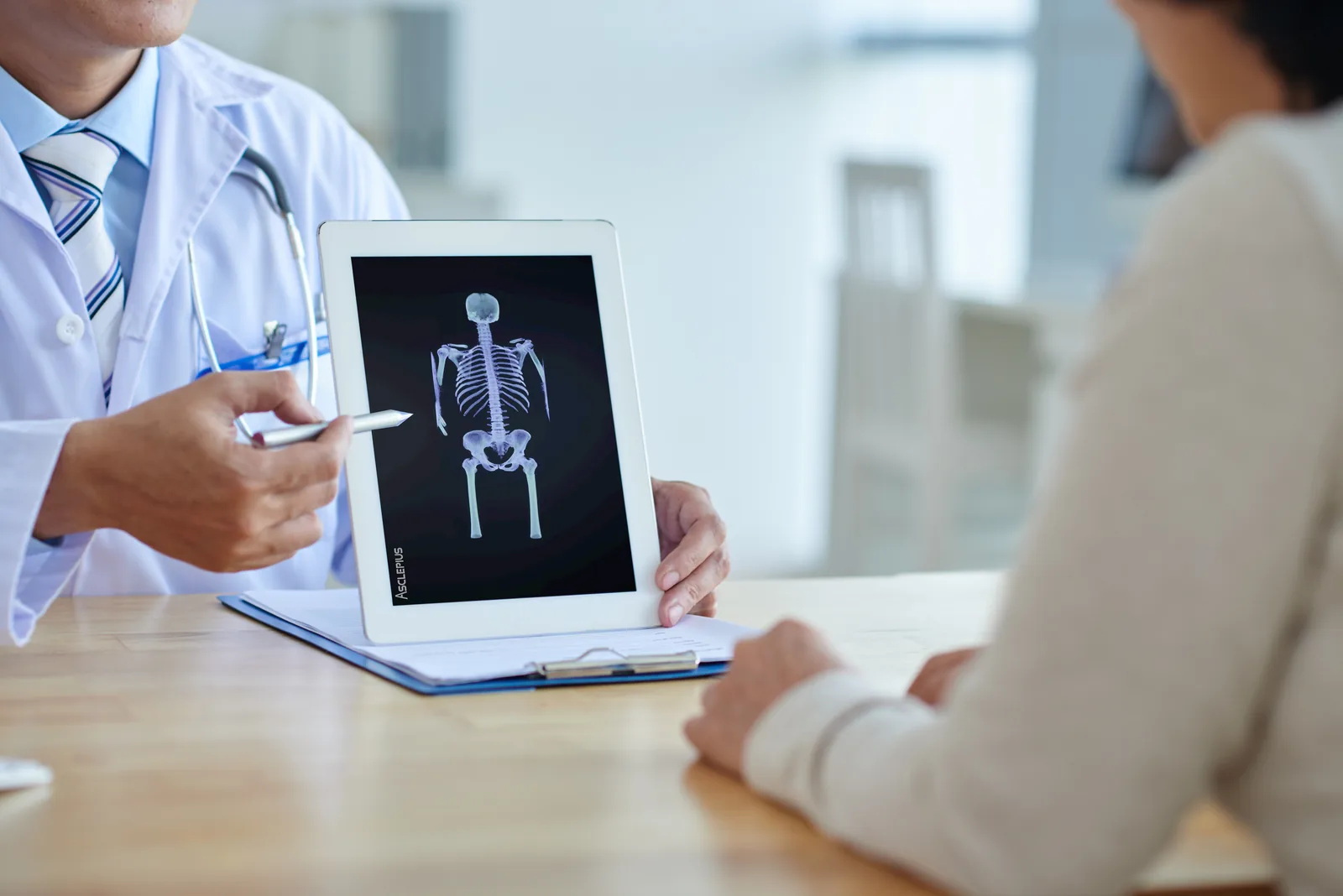
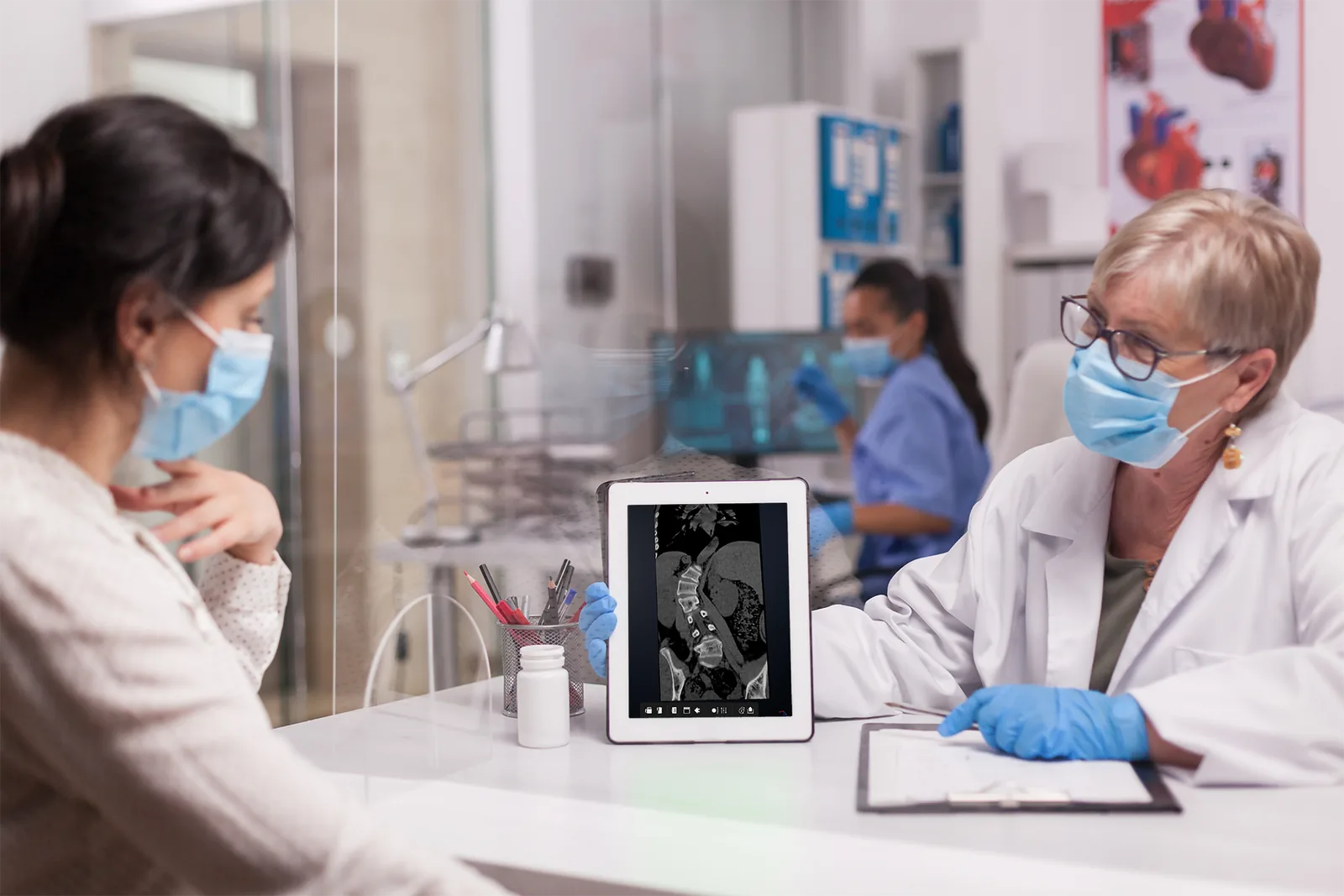
Visualised 3D imaging data
Detailed 3D anatomical images give the patient a clear, accurate view of their own structure and of any abnormality in it — which makes a diagnosis, a treatment plan and a surgical route far easier to take in.
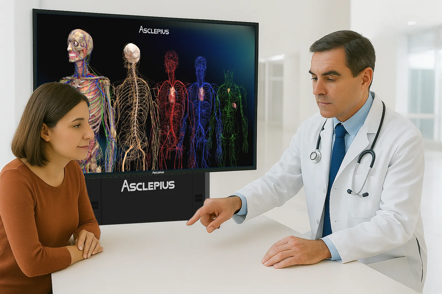
Interactive touchscreen simulation
The clinician walks the patient through the condition and the procedure on the touchscreen, showing exactly where and how it will be performed. It opens a dialogue rather than delivering a verdict.
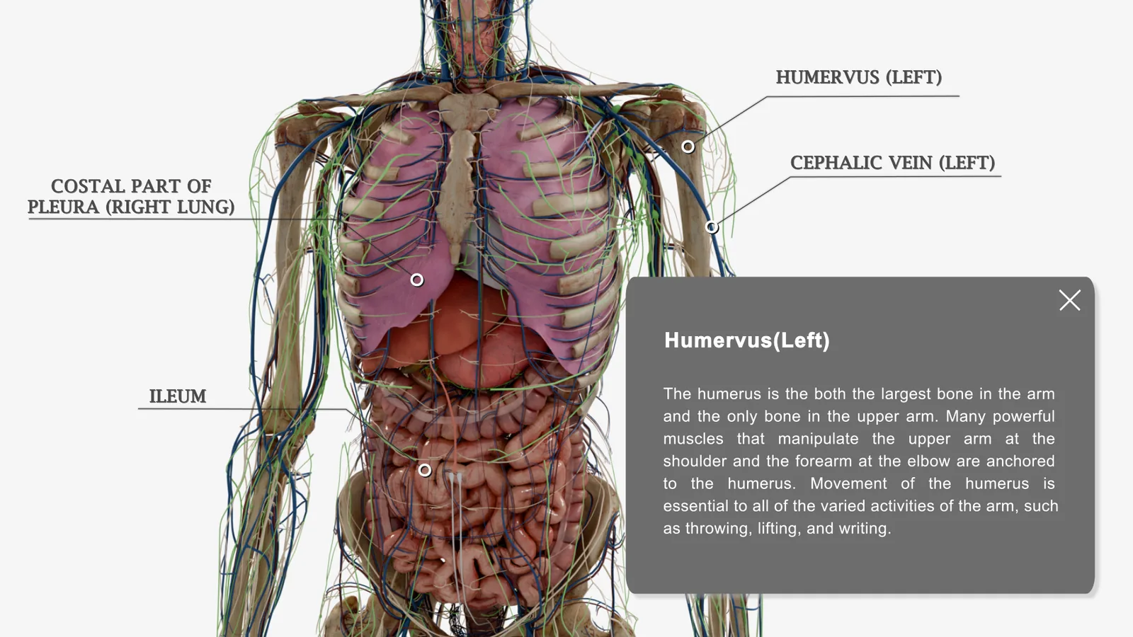
Complete anatomy, annotated
Every body region carries full anatomical data and detailed annotations, so both sides of the desk are working from the same explanation of what a structure is and what it does.
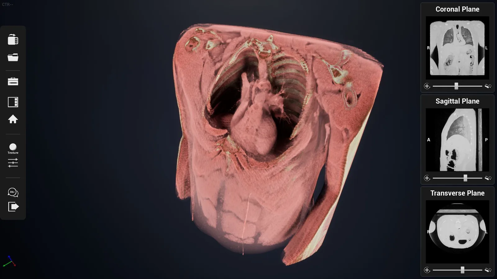
The patient's own CT and MRI
DICOM data goes straight in and overlays onto the 3D model, so the explanation is about this injury and this treatment rather than about a textbook case that merely resembles it.
Case Studies
What institutions reported afterwards
Universities, medical colleges, nursing programmes and secondary schools around the world have put Asclepius into anatomy teaching and measured what happened to their students' results. Six of those accounts are below.
Key benefits across the case studies
- Higher student scores — improvements of up to 30 % in student results, reported across a range of programmes
- More engagement — students find an interactive, hands-on session more absorbing than the traditional equivalent, and say so
- Better learning outcomes — from high-school biology through to advanced medical training, not only at one level
- Positive feedback from educators — who report the platform makes their own teaching more effective, not just more modern
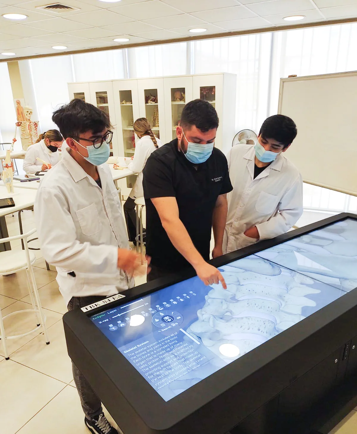
Anatomy education and exam scores
A university in the United States. Exam scores rose by up to 30 % after the table was adopted. Students reported understanding complex anatomical structures better through interactive 3D models and virtual dissection than through cadaver-based methods alone.
Radiology and imaging science
A medical college in the United Kingdom. Students on radiology and imaging science programmes saw an average 32 % improvement in GPA once Asclepius was part of the curriculum, having been able to visualise anatomical structures with far more precision.
Anatomy and physiology courses
A high school in Taiwan. Anatomy and physiology students averaged a 20 % improvement in exam scores. Interacting with 3D representations of the body made the harder physiological concepts easier to grasp.
High-school biology
A high school in the United States. Biology students recorded a 15 % increase in test scores, and reported greater engagement with the material once human anatomy could be explored in 3D.
Nursing and clinical training
A nursing college in Europe. Asclepius was built into clinical training programmes. Detailed visualisation of the body systems strengthened both theory and practical skill, and students showed more confidence in clinical settings.
Simulation labs and group work
A university in the United States. The table was used in simulation labs for group discussion and hands-on exploration. Being able to practise repeatedly, with no cadaver required, made it a popular fixture in the lab.
Figures are as reported to us by the institutions concerned. Names are withheld at their request.
Talk to an expert
Tell us how you would use it
A cadaver lab, a clinical training programme, a consulting room and a school science department each need a different model and a different software set. Tell us what the table would be doing and we will show you the combination that fits — online, or on site with the table in front of you.