
Foresee-X — Smart AR Surgical Glasses
See better. Operate smarter.
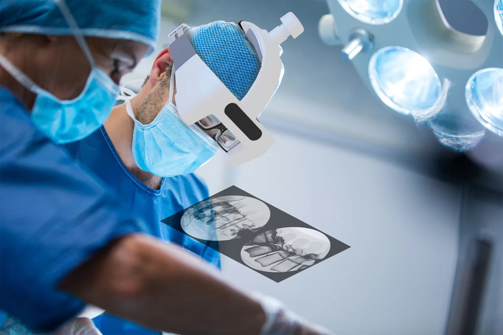
Foresee-X is a wearable augmented reality display system for image-guided procedures. It projects live medical images directly into the surgeon's field of view, so the picture arrives where the work is happening — and attention stops travelling between the patient and a monitor across the room.
CE marked · TFDA Class II licence MOHW-MD-No. 007801
What it is
What is Foresee-X?
Foresee-X is a wearable AR display system that integrates with the surgical imaging tools a theatre already runs — fluoroscopy, ultrasound, endoscopy and CT. It sends those images wirelessly to a lightweight headset, giving hands-free, heads-up visualisation for the length of the procedure.
The image goes where the surgeon is already looking
The clip alongside is recorded through the glasses themselves, in a real operating room. The endoscopic view sits as a panel in the upper part of the wearer's vision while their hands stay on the instruments and their eyes stay on the patient. Nothing tethers the surgeon to the equipment across the room, and no monitor has to be positioned for them to see — the display travels with them.
Images reach the headset over a 5G wireless link, and the same stream can be recorded or sent on for tele-mentoring and later review.
AR imaging in real time
2D and 3D medical images are displayed directly within the user's line of sight, live, for as long as the procedure runs.
Multi-device integration
Compatible with ultrasound, endoscopy, C-arm and O-arm imaging, among others — no equipment has to be replaced.
Wireless transmission and recording
Image streaming, recording and remote sharing, which makes tele-mentoring and post-case review part of the same workflow.
Lightweight and ergonomic
Built for comfort, clarity and low-latency visualisation, because the cases that need it most are the long ones.
Why AR
Why choose augmented reality?
Augmented reality changes where surgical information lives. With Foresee-X, live medical imaging is projected into the surgeon's field of view instead of onto a screen they have to turn towards — which is a change to precision, efficiency and patient safety, not just to ergonomics.
Real-time data access, without looking away
In a conventional theatre the surgeon repeatedly turns their head to check the external screens carrying the images that matter — X-ray, endoscope, ultrasound. Every one of those shifts costs time, and re-establishing the view of the surgical field afterwards is not free either. Foresee-X puts those same images in the line of sight, so the surgeon can keep working while they read them.
- Less head-turning and screen-glancing — the information is where the surgeon is already looking
- Better hand–eye coordination — the image and the instruments occupy the same view rather than opposite ends of the room
- Lower X-ray exposure — fewer unnecessary scans are taken when the existing image stays visible
- Support for teaching and remote collaboration — what the surgeon sees can be shared as it happens
Imaging beside the field

The live image sits alongside the operative field instead of on a monitor across the room — which matters most in complex and minimally invasive work, where the margin is smallest.
Effortless heads-up visualisation
Essential information stays accessible without looking away, which keeps the workflow moving and takes cognitive load off the surgeon in time-sensitive procedures.
Training and education
Trainees can observe a procedure from the operator's own perspective and receive step-by-step guidance, which is difficult to arrange any other way.
Better-informed decisions
Real-time imaging, a steadier workflow and fewer unnecessary scans together help the team decide faster and with more of the picture in front of them.
| Aspect | Foresee-X AR glasses | Traditional external monitor |
|---|---|---|
| Real-time image display | Images are projected directly into the surgeon's field of view | The surgeon turns their head towards an external monitor |
| External monitor required | No | Yes, and it is consulted frequently |
| Efficiency | No head turning, so focus and pace hold up through the case | Checking the screen needs head movement, which costs time |
| Precision | The image is aligned with the surgical site | Attention shifts between the patient and a display across the room |
| Focus and comfort | Attention stays on the patient, without the repeated look away | Focus is interrupted every time the surgeon looks away from the site |
| Collaboration | Up to two headsets can be worn at once, on the same live image | Limited, and feedback tends to arrive late |
The right-hand column describes the conventional monitor-based workflow that Foresee-X is designed to replace, not a competing product.
Multi-device integration
One headset, whatever you are looking through
Foresee-X is a display, not a replacement imaging system. It takes the signal from the equipment already standing in the room and puts it in front of the operator's eyes — which is why the same headset serves endoscopy, fluoroscopy and ultrasound-guided work without anything being swapped out.
Connects to what is already in the room
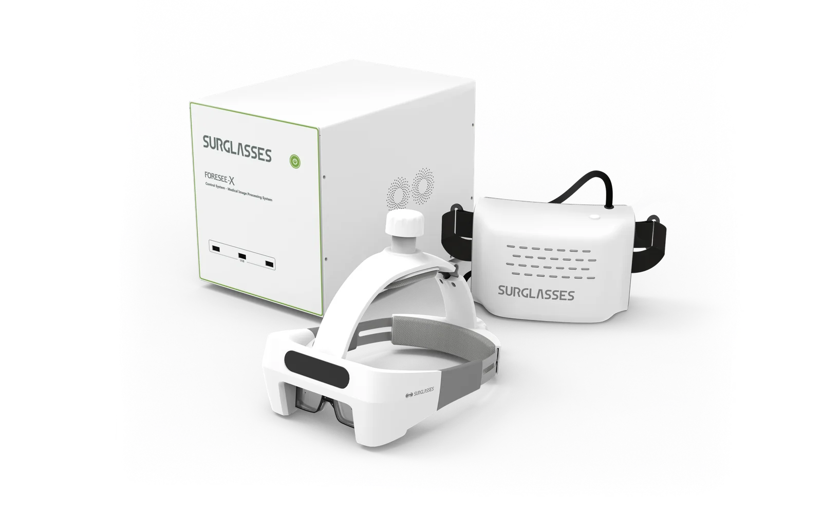
Existing imaging equipment connects to the Foresee-X control unit and its output goes to the headset from there. Nothing is fitted to the imaging device itself, nothing about the way it is used changes, and no external monitor has to be positioned for the surgeon any more.
Two imaging sources can be connected at the same time, with switching between them during the procedure — fluoroscopy and ultrasound in the same case, for example, without stopping to reconfigure anything.
Endoscopy
The endoscope view sits in the line of sight rather than on a tower across the room, which removes the constant glance to an external monitor. Fewer distractions, a more ergonomic visual position, and less fatigue in operations that run for hours.
Ultrasound
Overlaying the live ultrasound image supports quicker spatial recognition and more precise needle guidance, with no shifting of focus between the patient and a screen. Two headsets can be worn at once — operator and assistant, or trainee and mentor — on the same live image.
Clinical benefits
Precision and focus in the operating room
Foresee-X is more than an AR display. It is a surgical visualisation tool built to streamline the workflow around imaging, and to carry training and collaboration on the same equipment.
Real-time AR imaging, wirelessly
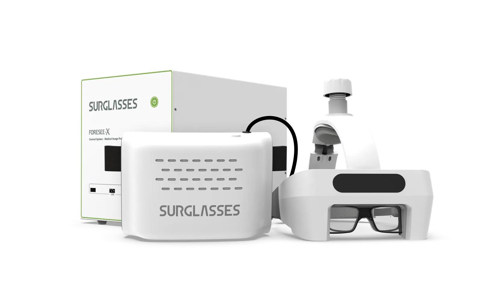
The system is three pieces: the headset, a small body-worn transmitter it plugs into, and a control unit that takes the feed from the imaging equipment and does the processing. The link between the transmitter and the control unit is wireless, so the surgeon carries everything they need and nothing runs back across the sterile field.
The heads-up display is continuous — it does not have to be summoned, and it does not go away while the surgeon's hands are occupied. The same link carries recording and remote sharing, so an observer outside the room sees exactly what the operator sees.
Enhanced surgical focus
Unnecessary head movement and screen-glancing are reduced, which helps surgeons and clinical staff hold their concentration across the whole procedure.
Dual-source switching
Two different imaging sources can be connected at once, with seamless switching between them — useful whenever a complex case needs more than one view.
Optimised visual environment
Colour, brightness and contrast are adjustable, so the display can be tuned to the operator's preference and to the lighting of the room they are working in.
Training and education ready
Suited to trainees, EMTs and remote observers: real-time visualisation supports hands-on learning and guidance given while the procedure is happening.
Clinical use cases
Where Foresee-X is used
Any procedure where the operator has to keep checking an image belongs on this list. The specialties below are the ones the system is used in today.
- Minimally invasive spine surgery — fluoroscopy stays in view through an approach made through a small working space
- Ultrasound-guided interventions — the probe image and the needle share one field of view
- Laparoscopic and thoracoscopic surgery — the scope view without the tower positioned for the surgeon's eyeline
- Emergency and trauma surgery — where time and attention are the scarcest things in the room
- Medical training and education — first-person observation and guidance, live or recorded
Regulatory status
CE marked and TFDA licensed
Foresee-X carries the CE mark as a Class I device under MDR (EU) 2017/745 and holds a Taiwan TFDA Class II medical device licence — Ministry of Health Medical Devices MOHW-MD-No. 007801. Availability and the approvals that apply vary by country; tell us where you are and we will confirm the position for your market.
Peer-reviewed
Backed by published clinical research
Foresee-X has been studied independently and the results are in the literature. In a pilot randomised crossover trial published in BMC Medical Education, 47 trainees across three Taiwanese hospitals placed central venous catheters using the Foresee-X system to display the ultrasound image, and again using a standard ultrasound monitor.
The trial's clearest finding was the one the technology is built around: the number of times an operator had to shift their gaze away from the patient fell from a median of five to one. Read the papers in full below.
Augmented reality visualization for ultrasound-guided interventions
A pilot randomized crossover trial to assess trainee performance and cognitive load. Liao S-C, Shao S-C, Gao S-Y, Lai EC-C. BMC Medical Education 2024;24:1058. Published 27 September 2024.
Read the trial (opens in a new tab)Augmented reality enhances medical education and improves efficiency
Liao S-C, Shao S-C, Gao S-Y, Lai EC-C. Medical Education, first published 14 February 2024.
Read the paper (opens in a new tab)Both papers are independent academic publications. They are linked here in full so the methods and limitations can be read alongside the results; the trial above describes itself as a pilot study.
Interested in Foresee-X?
Tell us what you are looking through
An endoscope, a C-arm or an ultrasound machine — whichever it is, we will show you how Foresee-X connects to it and what the view looks like from inside the headset. Online or in your own operating room or training programme.