
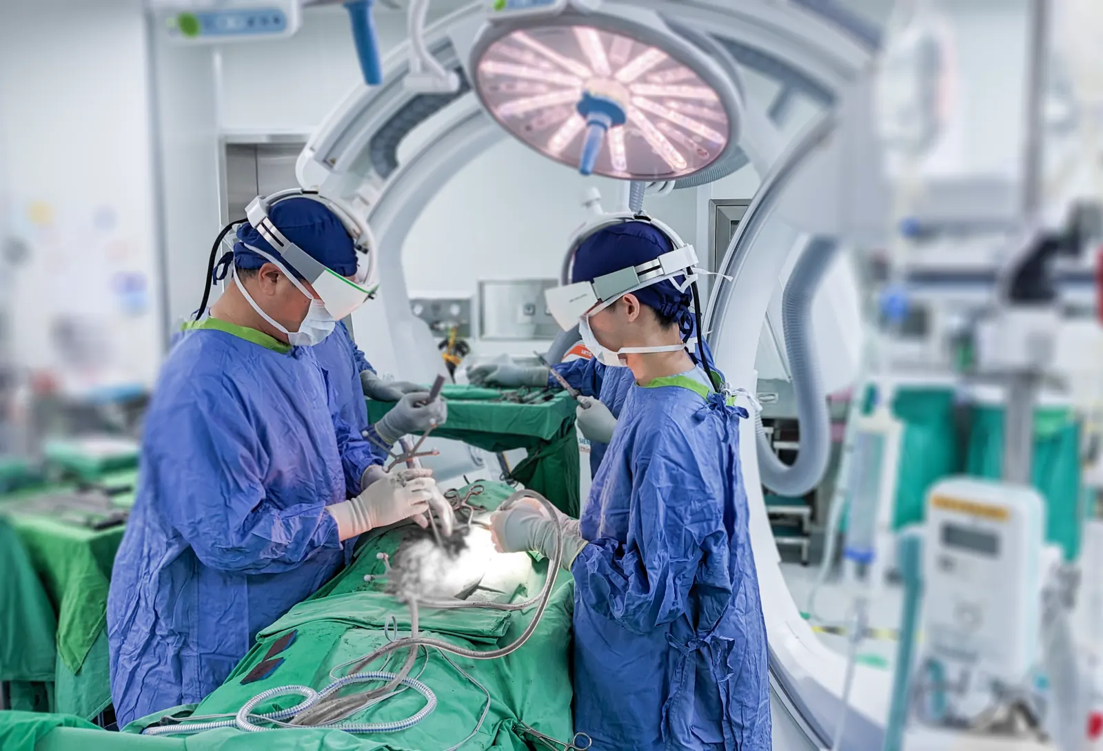
Caduceus S — Navigation
Spinal navigation, on the imaging you already own
Caduceus S is a 3D computer navigation system for spinal surgery. It reads the imaging your operating room already produces — a standard 2D C-arm, a 3D C-arm or an intraoperative CT — and turns it into live guidance, with a mean positional error under 2.0 mm in a cadaver study, without a preoperative CT scan and without a room full of extra equipment.
Licensed by Taiwan's TFDA as a medical device, MOHW-MD-No. 008401.
What it is
Why a spine surgeon needs navigation at all
Minimally invasive spinal surgery asks the surgeon to reach complex and delicate structures through a narrow field of view and a small working space. Navigation is what makes that reasonable: guided to the lesion directly, the surgeon does less damage to healthy tissue on the way in, and the accuracy of the approach stops depending on how much of the anatomy is exposed.
One system, whatever imaging is in the room
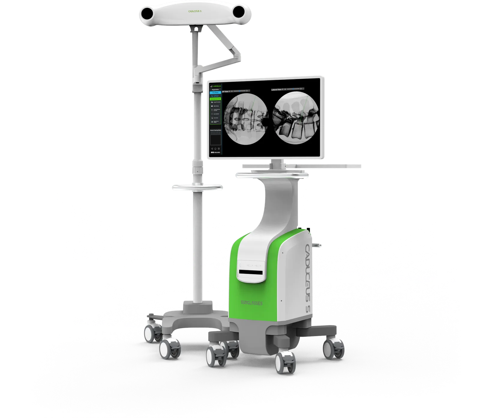
Caduceus S does not ask a hospital to replace its imaging. Three modules cover the three things a spine theatre is likely to already have — a plain 2D C-arm, a 3D C-arm, or an intraoperative CT — and each one registers the patient from images taken on that device. Nothing is bolted onto the existing equipment and the operating environment does not have to change.
Preoperative planning runs on the same workstation: load the DICOM data, simulate the approach beforehand, and have that plan on screen during the case.
Applicable across spinal implant surgery — scoliosis correction, spondylolisthesis, vertebral fracture and spinal fixation — as well as spinal tap procedures and minimally invasive spine surgery generally.
Precise positioning
The system reports the position of the instrument and its distance, direction and angle to the lesion throughout the case, with a mean positional error under 2.0 mm (cadaver study, FDA 510(k) K220554).
Minimally invasive by design
Locating the lesion quickly and confirming the instrument against it keeps the incision small — roughly 2 to 3 cm for a single segment, against the 5 to 10 cm an open approach needs.
Great compatibility
No auxiliary hardware has to be fitted to the existing imaging equipment, and the operating environment does not have to be rebuilt around the system.
Preoperative planning
DICOM data loaded into the navigation system supports simulation before the case, and that plan is displayed alongside the live imaging during it.
* Mean positional error: 1.80 mm in C-arm mode and 1.91 mm in CT mode; cadaver study, FDA 510(k) K220554.
Three configurations
Pick the module that matches your imaging
The three modules are the same navigation system reading three different sources. What changes between them is how the patient is registered, how much X-ray the case costs, and which kind of operating room they suit. All three work with the Caduceus S AR glasses.
| Aspect | 2D C-arm module | 3D C-arm module | Intraoperative CT module |
|---|---|---|---|
| Imaging source | A standard 2D C-arm | A 3D C-arm | An O-arm intraoperative CT |
| Registration | Simplified registration from the C-arm images themselves | Automatic, once the 3D scan is complete | Automatic, once the CT scan is complete |
| X-ray images taken | About 2 to 4 for the whole case | No further C-arm shots after the scan | No further C-arm shots after the scan |
| Equipment compatibility | A wide range of C-arm brands, using what is already in the room | Various registration tools; DICOM loaded straight into the workstation | O-arm, with no extra connections or configuration |
| Best suited to | Outpatient and ambulatory surgery centres already running a C-arm | Complex cases — four or more vertebrae, small curvatures or rotation | Mid- to high-end operating rooms, including deformity such as scoliosis |
| Works with the AR glasses | Yes | Yes | Yes |
All three columns are Caduceus S configurations, not competing products. Which one fits is a question about the imaging you already own, not about which is better.
Module 01
2D C-arm module
The 2D C-arm module runs spinal navigation from plain 2D fluoroscopy. It connects to a wide range of C-arm equipment already in use, which is what makes navigation possible in rooms where it has traditionally been out of reach.
Navigation with just a standard C-arm
Outpatient and ambulatory surgery centres run a great deal of spinal surgery, and most of them do it without navigation — not because they do not want it, but because their equipment will not carry it. This module removes that barrier: no complex setup, no additional imaging device, accurate guidance from the C-arm that is already parked in the room.
- Supports multiple C-arm brands — existing equipment is used as it stands, with no additional purchase and no complex configuration
- Fast and easy operation — a straightforward interface that fits the current workflow, so the team starts navigating without a long learning curve
- Simplified imaging process — registration is quicker, which takes time out of the setup and out of the case
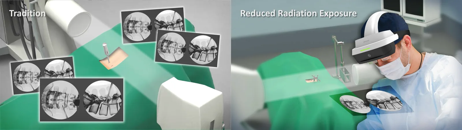
- Reduced radiation exposure — fewer C-arm shots are needed, which is safer for the patient and for everyone standing in the room
Module 02
3D C-arm module
Where the anatomy is complicated, a 3D volume beats two flat views. This module takes the 3D C-arm scan, registers it automatically and drives navigation from it — which is what makes it the right choice for four or more levels, small curvatures, or a rotated vertebra.
Navigating on the 3D volume
The reconstructed volume, the planned trajectory and the instrument position sit on one screen, so the approach is checked against the anatomy rather than inferred from two projections.
Quick 3D image conversion
The scan is converted and brought into the workflow fast enough that it does not interrupt the procedure it is meant to guide.
Registration, simplified
Various registration tools are supported, and DICOM images load straight into the navigation workstation — so the case continues without going back for more C-arm imaging.
Fast conversion
3D images are converted efficiently and folded into the surgeon's workflow with minimal disruption to the procedure.
Simplified registration
The system supports a range of registration tools and integrates smoothly with the 3D C-arm, with DICOM loaded directly into the navigation workstation.
Automatic registration
Once the 3D scan finishes, the software registers the images itself — removing the traditional C-arm shots and the extra imaging that goes with them.
Built for complex cases
Four or more vertebrae, small curvatures or rotations such as L3 and L4 — the cases where precise navigation matters most.
Module 03
Intraoperative CT module
The intraoperative CT module pairs Caduceus S with an O-arm. The scan is taken in theatre, on the patient as they are positioned, and the navigation runs on that — real-time imaging with the accuracy of CT, and the surgeon's attention still on the patient.
Automatic registration from the scan
The O-arm captures a real-time CT of the surgical area and the system registers it itself. No additional C-arm imaging, and less time spent acquiring images at all.
No excess equipment
Nothing else has to be connected or configured. 3D imaging joins the surgical workflow rather than being bolted onto the side of it.
For mid- to high-end theatres
Suited to a range of surgical settings, from mid-level operating rooms up to the most advanced facilities.
Precision where anatomy is deformed
Particularly useful in complex spinal work such as scoliosis, where the margin for error is smallest; X-ray images can be brought into the navigation alongside the CT.
Low radiation
Two to four images, and no CT beforehand
Traditional spinal surgery tends to take a CT before the operation and more imaging during it. Caduceus S needs neither: the initial 2 to 4 X-ray images taken in theatre are enough to navigate a minimally invasive case.
Why the image count matters
Radiation dose is cumulative, and the long-term risk it carries rises with it, which is why imaging that is not needed should not be taken. For scale: local authorities cap the dose a member of the public should receive in a year at 5 millisieverts, and a single CT scan is roughly 1.5 to 2 millisieverts of that budget — before the patient has reached the operating table at all.
- No preoperative CT — the scan that would normally be taken before surgery is not required, so its dose is never delivered
- No additional intraoperative CT — the whole case can be navigated from the C-arm images already taken
- A shorter route to theatre — after the outpatient consultation the patient can go straight to the operating room, with no separate appointment for a preoperative scan
Optional integration
Add the AR glasses, and the monitor stops mattering
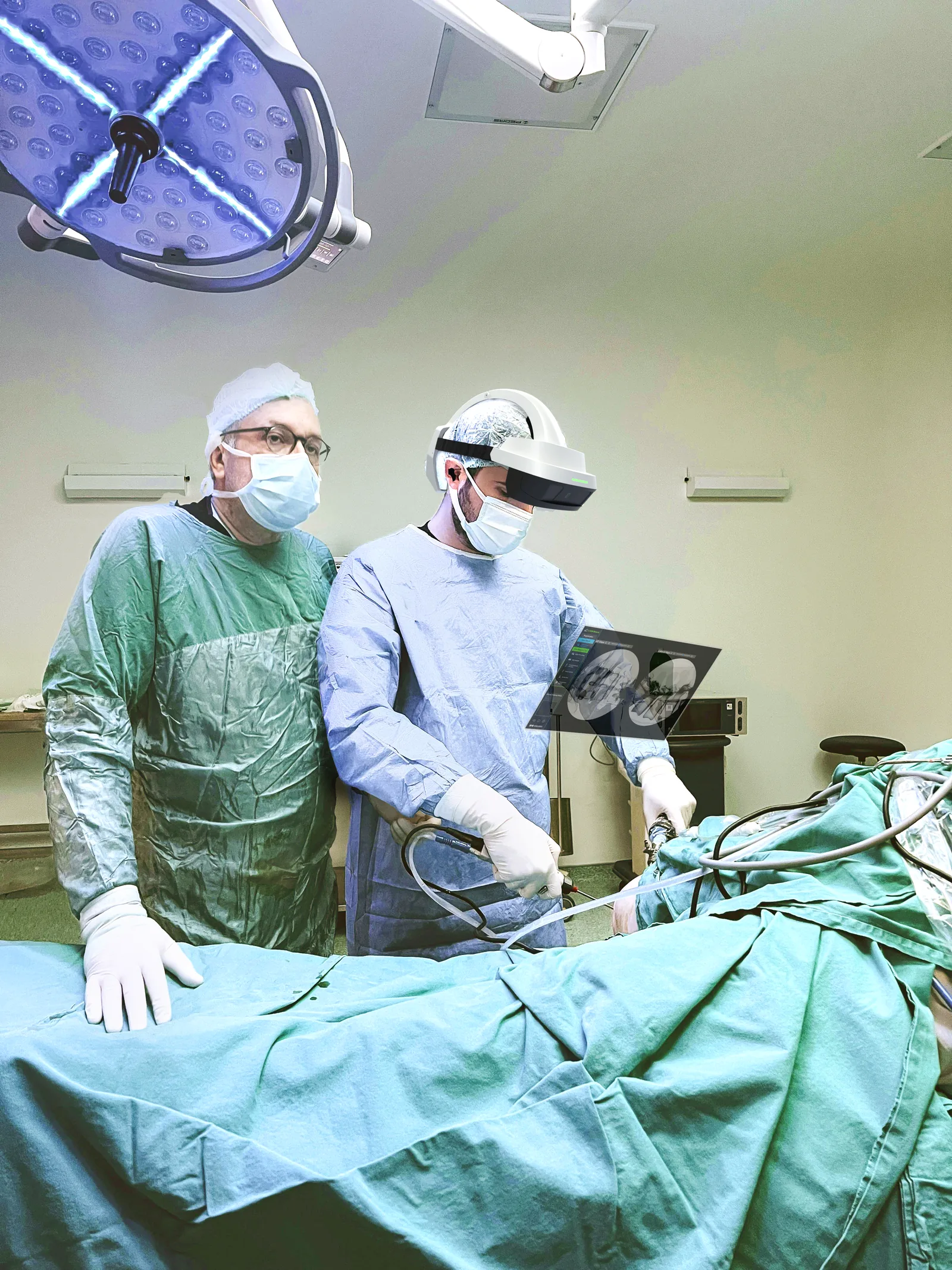
Every one of the three modules can be paired with the Caduceus S AR glasses. The navigation image then appears in the surgeon's own field of view instead of on a screen across the room — the same guidance, without the glance away from the wound.
The same navigation, in the line of sight
- No need to turn their heads — critical navigation data is displayed where the surgeon is already looking, so attention stays on the patient
- Real-time 3D superimposition — with the 3D C-arm and intraoperative CT modules the volume is laid over the patient as they lie on the table, which is where complex navigation is hardest to do from a screen
Navigation that fits the room you have
Tell us what imaging you already own
A 2D C-arm, a 3D C-arm or an intraoperative CT — whichever it is, there is a Caduceus S module that runs on it. Tell us what is in your theatre and the procedures you do most often, and we will show you the configuration that fits, online or on site.