
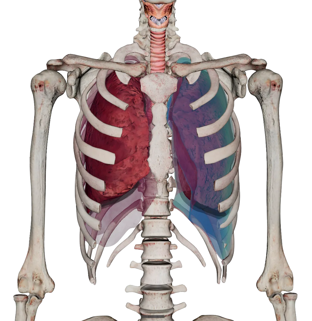
Asclepius Software
Eight modules, one dissection table
Six software packages run on the Asclepius table — Anatomy, Animal Anatomy, Embryology, Histopathology Atlas, OOOPDS Radiology and CTRender — and two more come with it: an online examination platform and a Windows companion app. This page goes through what is inside each one.
Module 01 — Anatomy
A next-generation platform for digital human dissection
The core package, and the reason most institutions buy the table. One of the most comprehensive digital anatomical libraries available today: every organ, vessel, nerve and system modelled and labelled individually, on bodies a class can take apart as many times as it needs to.
- 5,000+ physiological structures — meticulously modelled and labelled, each one separable from the layers around it
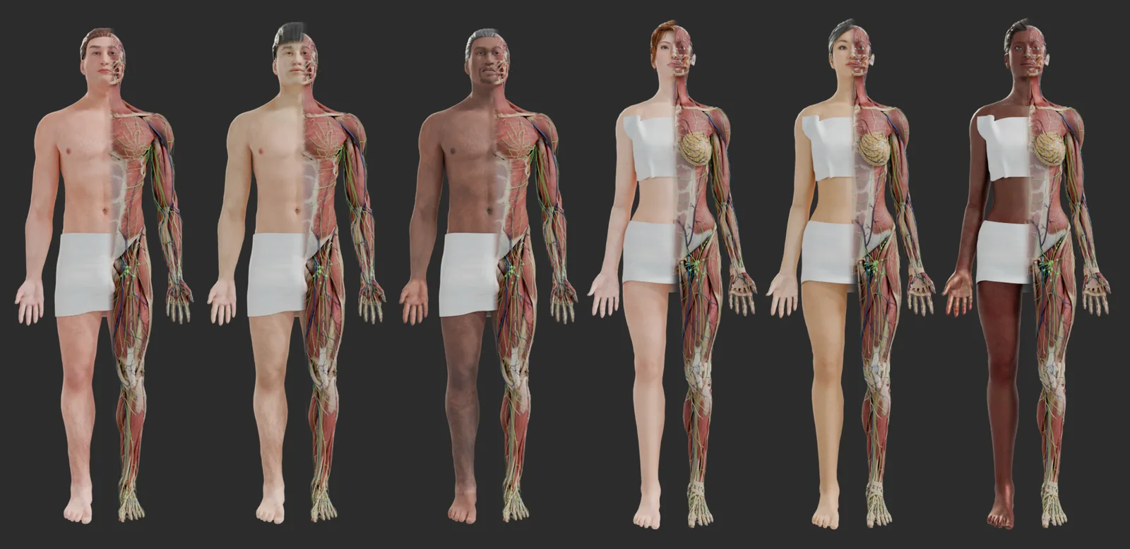
- 6 full-body cadaver models — male and female bodies from diverse ethnic backgrounds, so anatomical variation across populations can be taught rather than assumed
- 912 regional anatomy modules — categorised for targeted teaching, from head and neck through the limbs to the internal organs
- 75 real cadaver image comparisons — each structure paired with a photograph of the real specimen, so the digital model can be checked against the thing itself
- 5 endoscopic simulations — the digestive, respiratory and reproductive systems explored with interactive 3D endoscopes, alongside reference video from real procedures
Teaching tools built around the model
The library is only half of it. Everything below is a tool an instructor uses live, in front of a group, without consuming anything that has to be replaced afterwards.
- Layer-by-layer dissection and free cutting — draw any cutting path you like, from a single organ to a full-body section, and examine how the systems sit against each other
- Sectional anatomy from real frozen slices — coronal, sagittal and transverse planes built from actual cross-sectional body slices
- Interactive labelling — tap any structure for its name and description, isolate it, or hide everything around it
- Virtual endoscope — travel the large intestine, digestive tract, respiratory system and vaginal canal, each with real endoscopic video for reference
- Animated tutorials and physiological simulation — annotated heart models with a realistic heartbeat and section views, with more animated content added over time
- Brush tool for educators — draw, mark and write over the model during a lecture, then save the screenshot to USB for the next class
Module 02 — Animal Anatomy
Forty species, dissected the same way as the human models
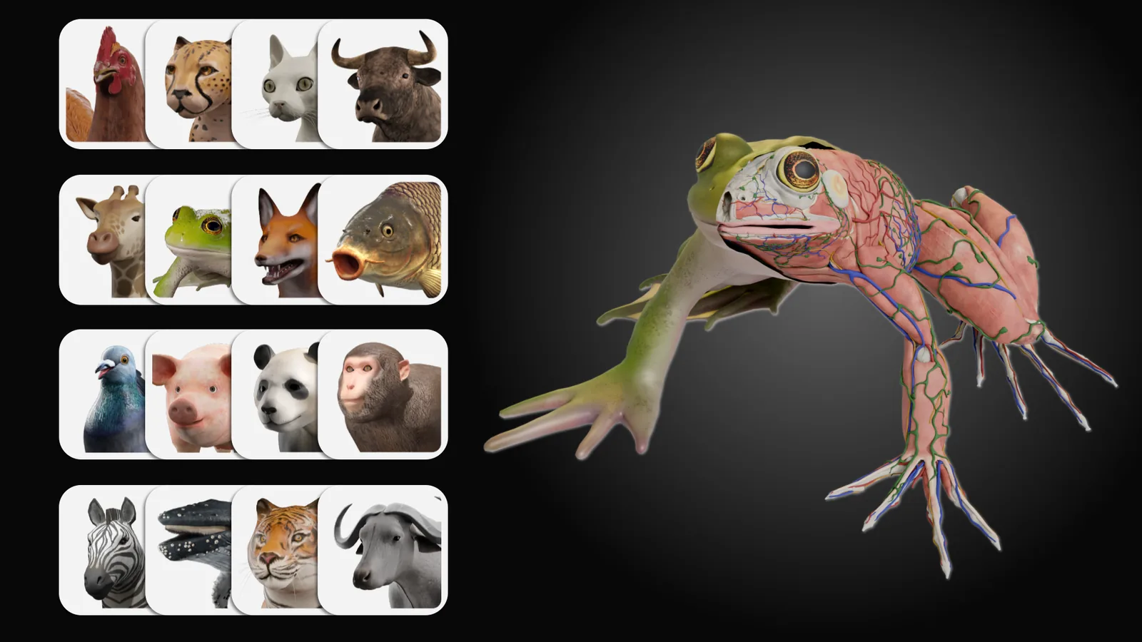
Comparative anatomy without a specimen. Over forty animals are modelled in 3D to the same standard as the human bodies, so veterinary, biology and zoology programmes can dissect repeatedly, safely and without a supply chain.
- Over 40 animal species — detailed 3D models spanning mammals, reptiles, birds and fish, so structures can be compared across species
- Every structure is interactive — click an organ, system or tissue for its name and a full explanation of what it does and where it sits
- Unlimited layered dissection — work through a body layer by layer at your own pace, internal and external, as often as the class needs
- Custom models on request — if a programme needs a species or a level of detail that is not in the library, Surglasses will build it
Module 03 — Embryology
The whole journey from conception to birth, in 3D
An interactive study of human development that a textbook cannot match: the embryo and fetus modelled at every stage, the pregnant body around it, and the abnormalities that a clinician will eventually have to recognise.
- Carnegie stages 7, 9 and 11, then week 5 to week 42 — a detailed model for every week of gestation, from early neural development to the final stages before birth
- Pregnant female anatomy — the anatomical changes of pregnancy, plus a simulation of the blood exchange between mother and embryo and the function of the placenta
- 100+ embryonic abnormality case studies — genetic conditions, birth defects and developmental disorders, each with an explanation and a visual simulation
- Unlimited layered dissection — every anatomical and physiological structure of both the embryo and the pregnant woman, taken apart layer by layer
Embryology is one of the two optional packages. It is included as standard on the Asclepius AI Table.
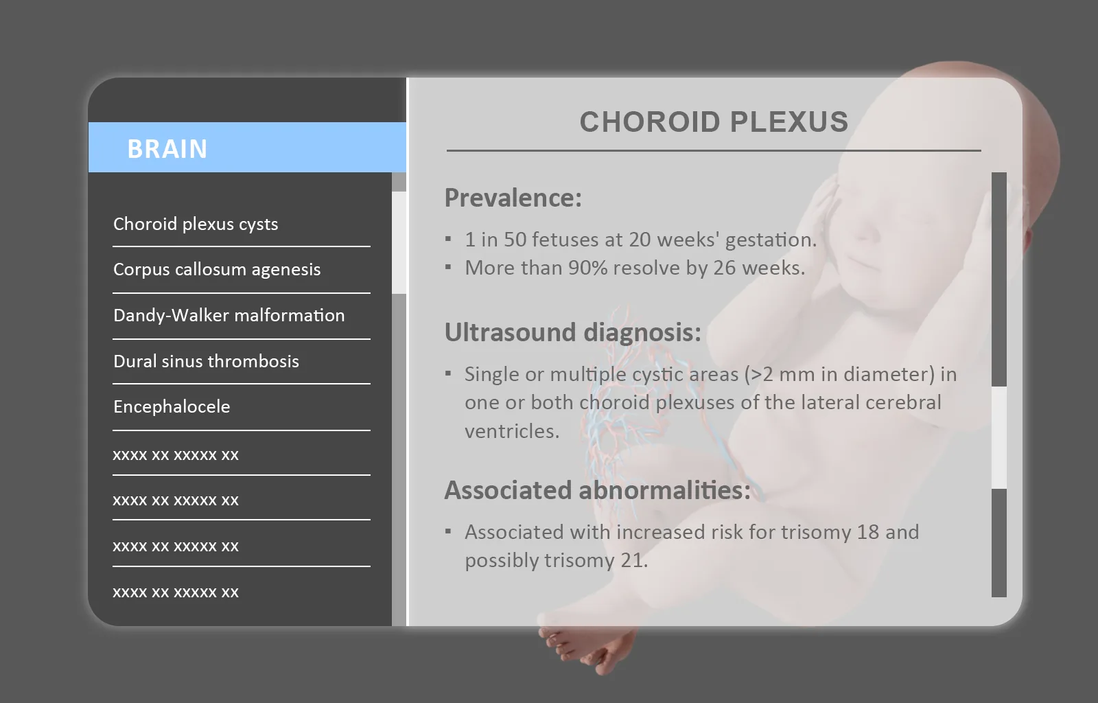
Week by week, in the round
Each week of gestation is a model in its own right, not a plate on a page — so the change from one week to the next can be shown from any angle.
Blood exchange, simulated
The circulation between mother and embryo running in place, so placental function can be taught as a process rather than described as one.
Cut anywhere, peel anything
The same free cutting and layer removal as the anatomy package, applied to the embryo and to the body carrying it.
Module 04 — Histopathology Atlas
A thousand digital slides, sharp down to the cell
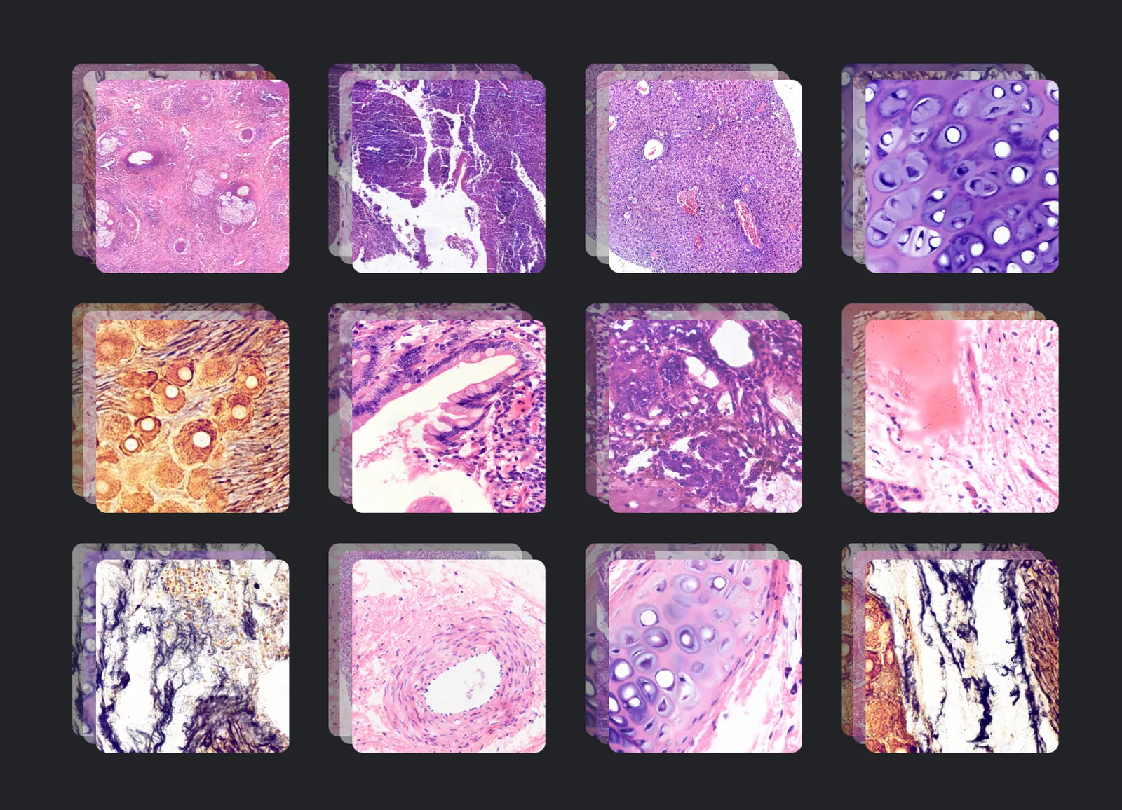
A digital histology and pathology slide viewer built into the table. Instead of one student at a microscope, the whole group looks at the same slide at once — and the slide never degrades, gets misplaced or has to be re-cut.
- 1,000+ histology and pathology slides — a library covering the range of human tissue types and pathological conditions, each one digitised to preserve microscopic accuracy
- High-resolution imaging — an average of 10,000 × 10,000 pixels per slide, so magnifying costs no clarity
- Expert annotations — every slide carries a written description of the key features to identify and what they mean
- Zoom to any magnification — in and out instantly, under the viewer's control rather than the objective turret's
- Image adjustment tools — brightness, contrast and sharpness, to bring out the structure being taught
- Import your own slides — teachers can load their own virtual slide files, so departmental material sits alongside the built-in library
Module 05 — OOOPDS Radiology
Real clinical imaging, from DICOM to a 3D object
OOOPDS Radiology puts a library of real clinical studies in front of students, with the tools to segment them, measure them, turn them into three-dimensional objects and plan treatment on them — the same core imaging workflow used in clinical practice, scaled for a classroom.
The module is built on OOOPDS, our medical image processing software, which is cleared by the US FDA under 510(k) K254214 for image segmentation, measurement and treatment planning. OOOPDS for surgical planning
- 1,200+ real clinical DICOM CT/MRI cases — selected for range and image quality, so the examples in front of a class are the ones they will meet in practice
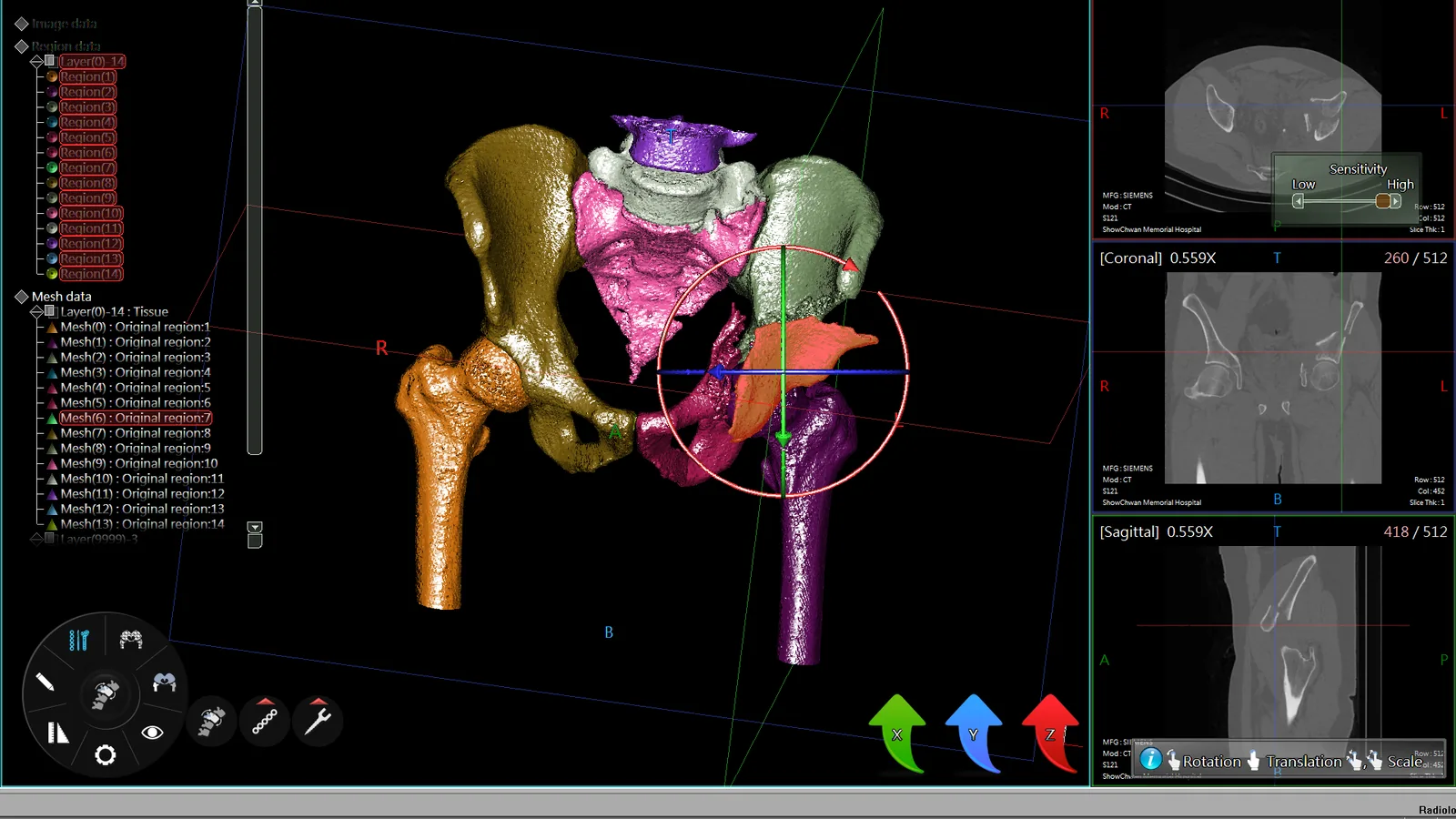
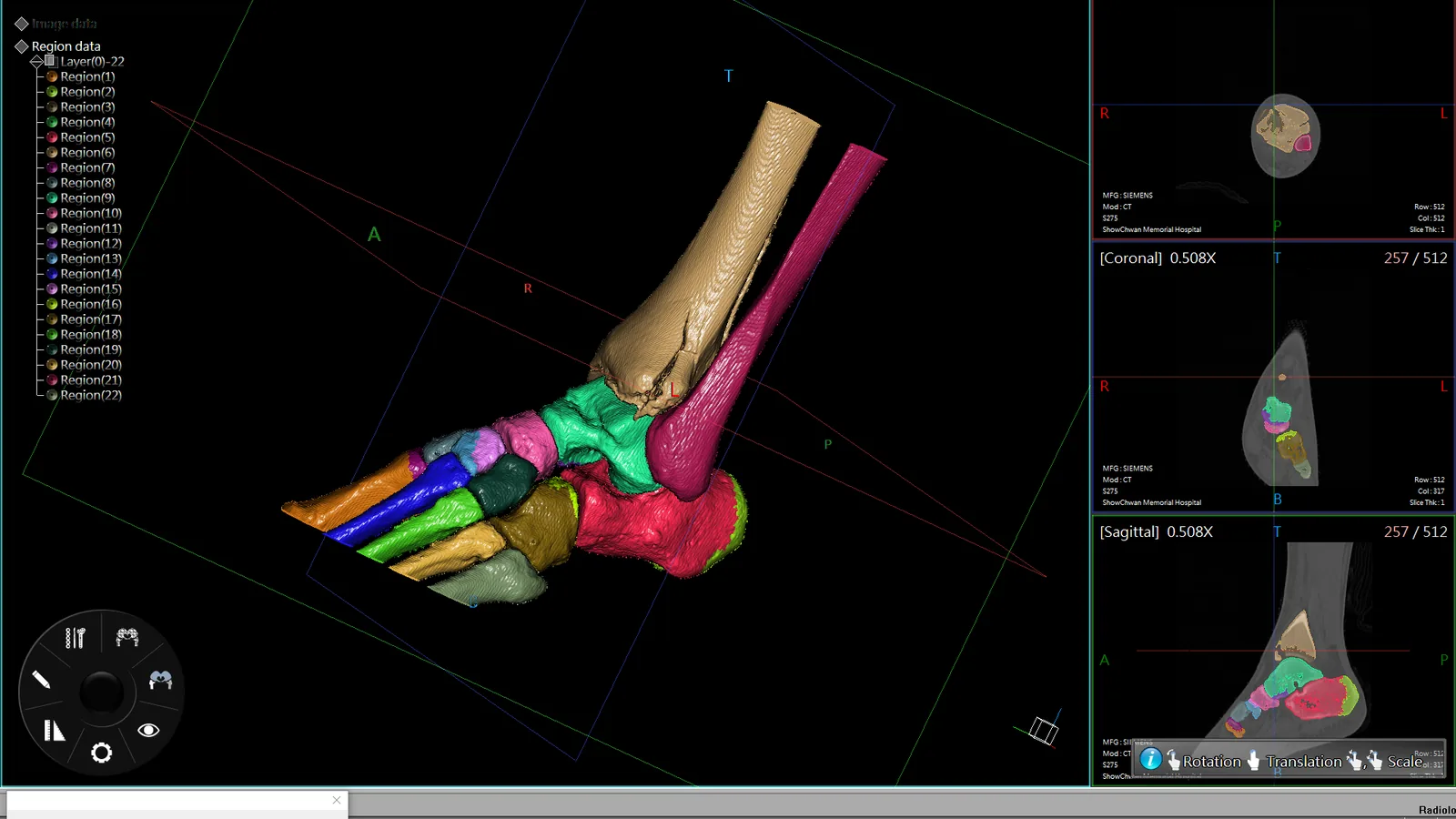
- Semi-automatic bone segmentation — extract precise anatomical regions out of complex CT and MRI data without tracing every slice
- 3D image to 3D model in seconds — and an export in a format suitable for 3D printing, so the study can leave the screen
- Pre-surgical planning simulation — customised bone plates and screws, spinal puncture and dental implant procedures, planned before they are performed
- Advanced measurement tools — measure and analyse the anatomical structures in a study, which is where diagnostic judgement actually gets built
- Load your own DICOM data — teach from your own hospital's cases, and integrate with PACS
Segment the bone, not the noise
Semi-automatic segmentation pulls each bone out of the study as its own region, so a complex joint can be taken apart and rebuilt.
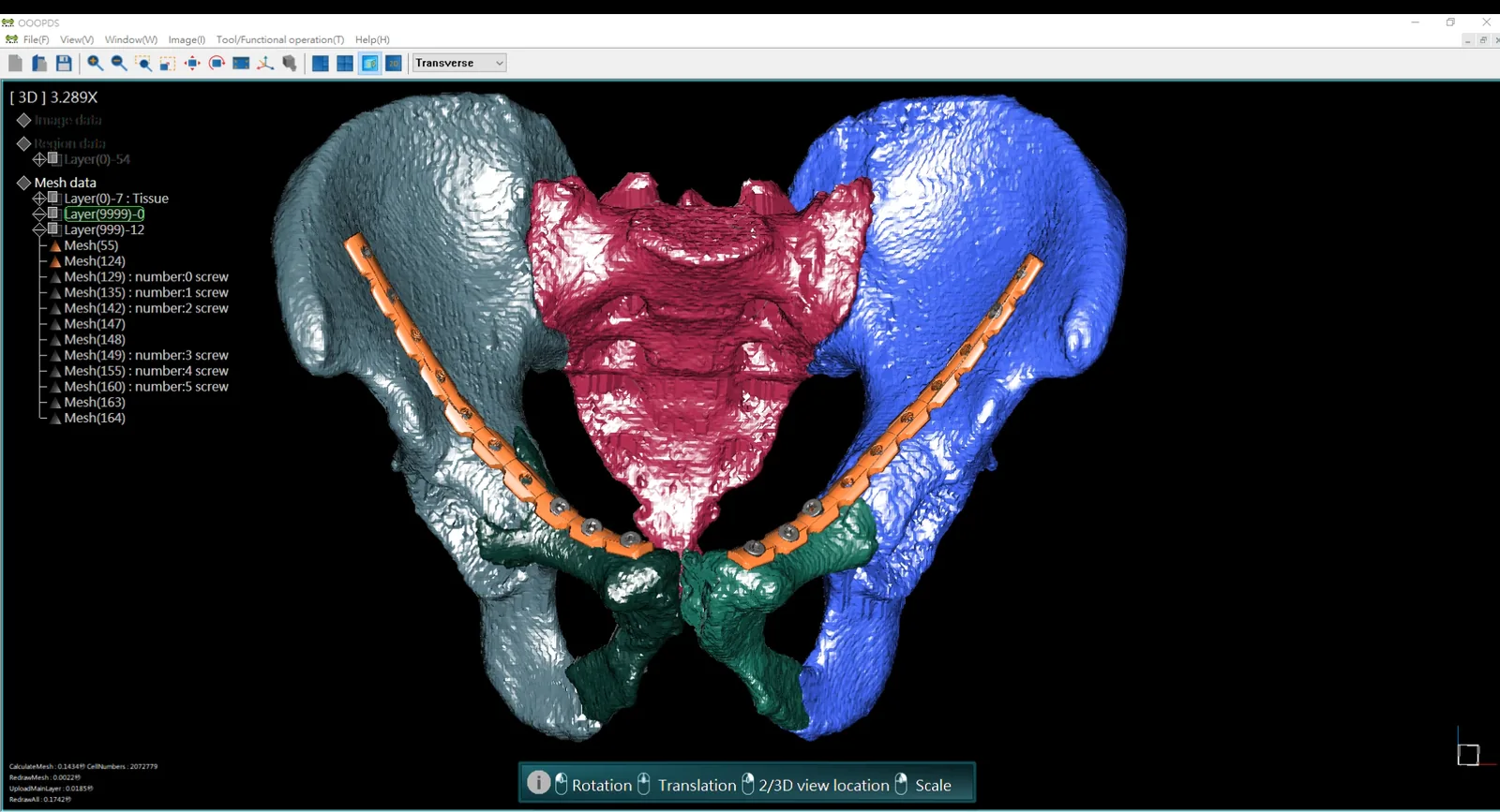
Plan the plate before the operation
Customised bone plates and screws are shaped against the patient's own anatomy, as an exercise students can work through and compare.
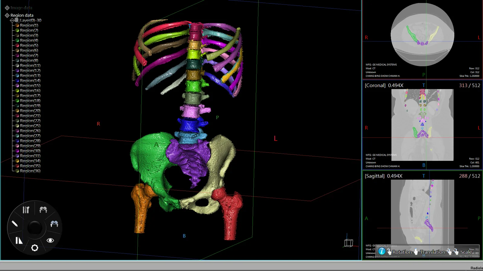
A library of real studies
Over 1,200 clinical CT and MRI cases are on the table already, and a department can load its own alongside them.
Module 06 — CTRender
Turn a CT study into something the class can turn over
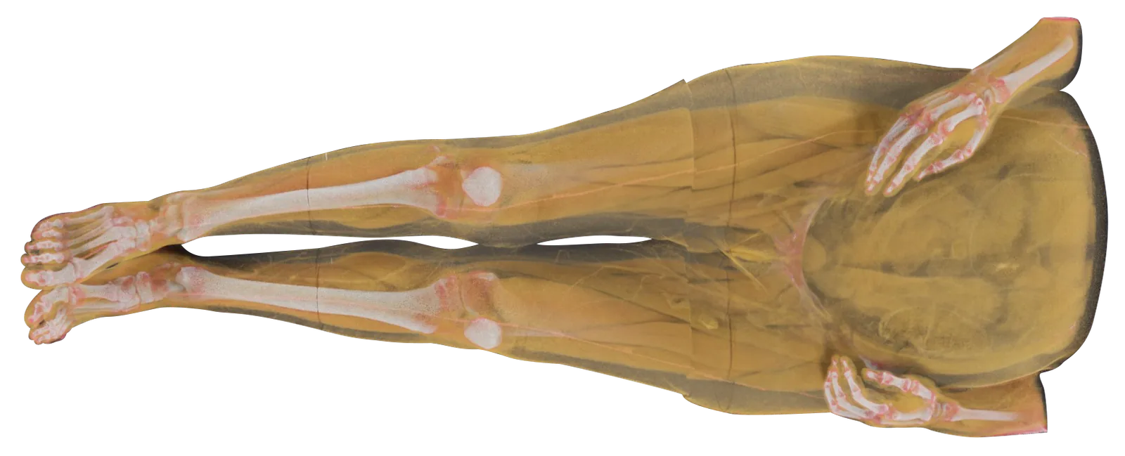
CTRender takes DICOM CT and MRI data and renders it into a vivid, interactive 3D volume. Where OOOPDS Radiology is about analysis and planning, CTRender is about seeing — what the tissue looks like, where it sits, and how a slice relates to the body it came from.
- Preset rendering modes — soft tissue (muscle, fat, vessels) and hard tissue (bone, joints) separated by colour and texture, with every parameter open to tuning
- Transparency, contrast and colour layers — bring one structure forward and let the rest fall back, without leaving the volume
- Multi-axial dissection and slicing — section the volume in real time along any plane, or cut through it on the standard axial, coronal and sagittal axes
- CT and 3D side by side — compare a slice with its rendering, next to each other or superimposed, and switch between the 2D radiological view and the 3D anatomical one
Module 07 — Online Examination
Set the test, mark it automatically, see who is struggling
The online examination platform is included with the Asclepius table at no extra cost. Instructors build assessments that match what they actually taught; students get their result the moment they finish; and the department gets a record of how the cohort is doing.
- A question bank for nursing and healthcare examinations — drawn from the professional licensure examinations of several countries, covering a broad range of topics and practical scenarios
- Customisable exam content — set the topics, the difficulty and the time limit so the assessment matches the curriculum rather than the other way round
- Real-time grading and feedback — students see where they went wrong straight away, while the question is still fresh
- Assessment by subject and module — anatomy, physiology or clinical skills, assigned alongside the course it belongs to
- Grouping and scheduling — organise students by where they are in the programme and give each group the assessment that fits
- Detailed progress reports — monitor a cohort, spot the trend rather than the individual mark, and adjust teaching accordingly
Separate sign-in for teachers and students. Students register by email, instructors approve them and release the questions with a time limit, and both sides see the result as soon as it is submitted.
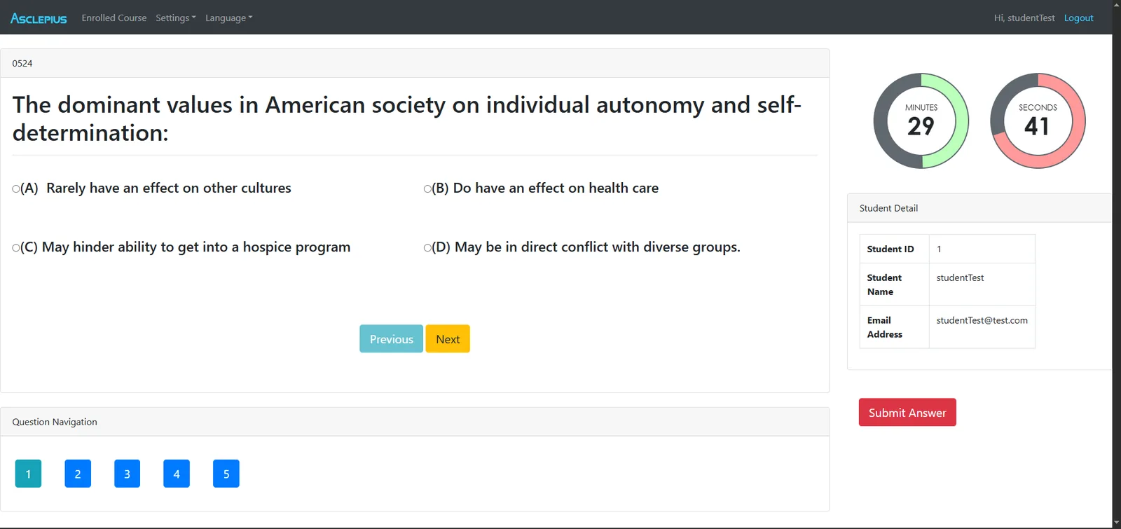
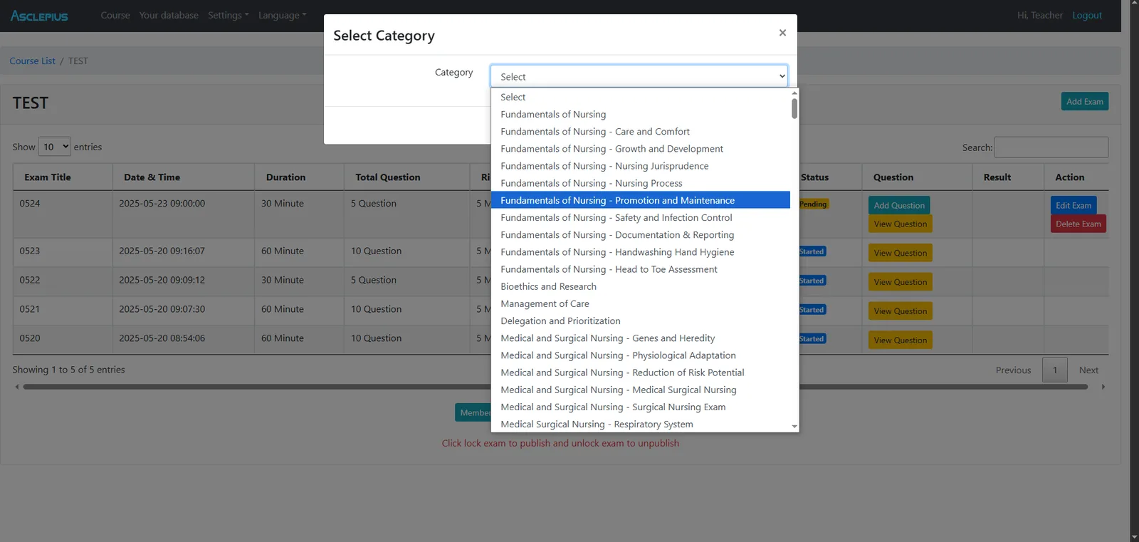
Assessments by subject
Exams are created against a named subject area, so a test can be pointed at exactly the part of the course that needs checking.
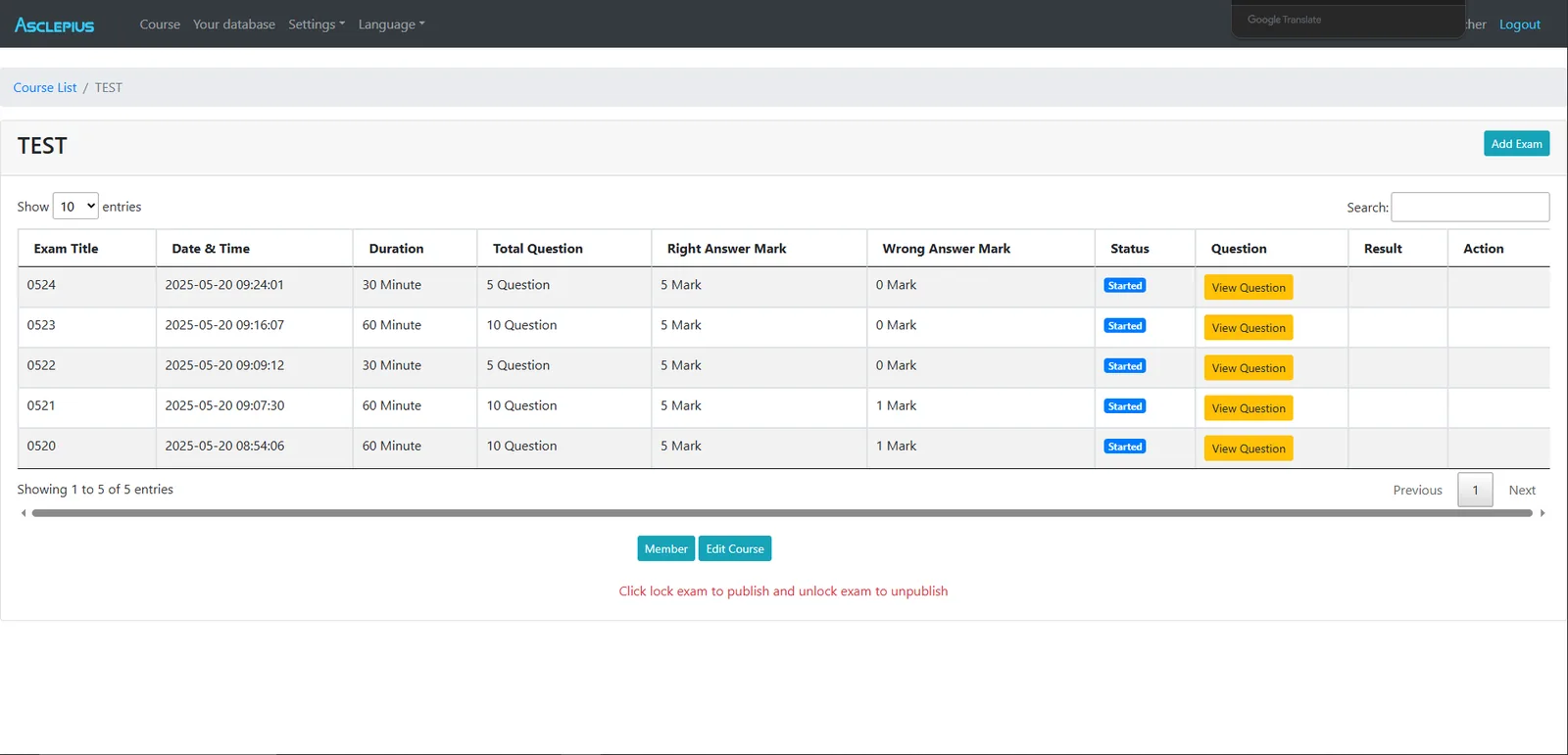
Build a quiz mid-lecture
An exam can be assembled, published and collected inside one class, with every student's score gathered automatically.
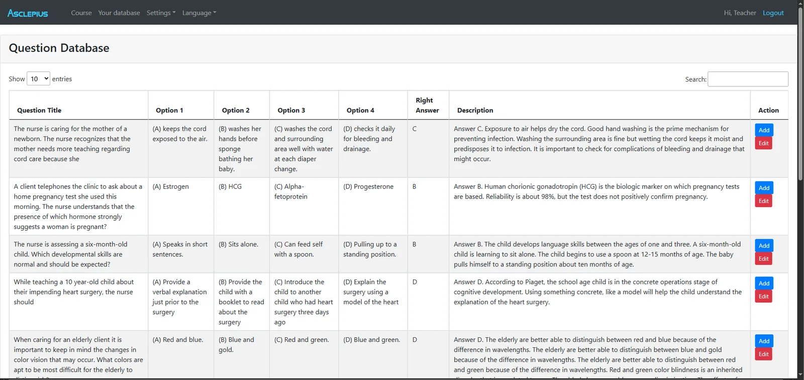
Write your own questions
Departmental questions sit alongside the supplied bank, so an institution's own material is not a separate system to maintain.
The table itself also carries a built-in offline examination system with its own question bank and several question formats, so students can test themselves at the table with no internet connection at all.
Module 08 — Windows App
Asclepius Anatomy Online — the same anatomy on a Windows PC
Available with the purchase of an Asclepius table, the Windows app gives educators and students a simple, interactive way to teach and explore human anatomy away from the table itself — in a seminar room, a library, or at home the night before the lab.
- Comprehensive human anatomy — the full anatomical structures with their physiological terms, explanations and context
- Layered anatomy systems — work outwards from bone through muscle, the circulatory system and the rest, one layer at a time
- Regional anatomy — study one area of the body in depth rather than the whole thing at once
- A fully interactive dissection — the same digital dissection experience as the table, on a desktop screen
The Windows app is available exclusively to institutions that own an Asclepius table. It is optional and is purchased separately.
See the software running
Bring us the course, we will show you the modules
Tell us what you teach and to whom, and we will run through the packages that actually matter for it — which are included, which are optional, and what a session on the table looks like for your students.