
Caduceus S — AR Navigation
Navigation you never look away from
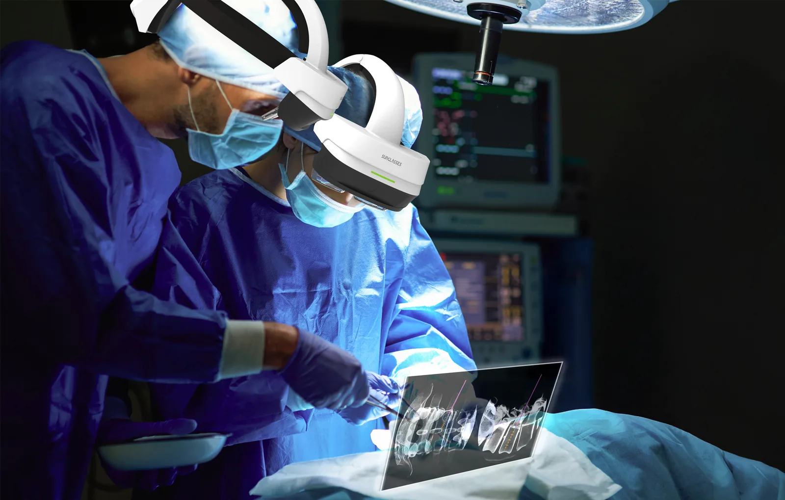
Caduceus S is an augmented reality navigation system for spinal surgery. It projects the navigation image into the surgeon's own line of sight, so the plan, the instrument and the anatomy stay in one view — no glance across the room at a monitor, no preoperative CT, and two to four fluoroscopy images before the first screw goes in.
Cleared by the US FDA under 510(k), and licensed as a Class II medical device by Taiwan's TFDA and the Thai FDA.
What it is
Augmented reality, brought into the surgical field
Caduceus S is an advanced augmented reality surgical navigation system built for spinal procedures. By projecting real-time AR visuals directly into the surgeon's field of view, it supports precise navigation and minimally invasive technique without adding a screen, a scan or a second imaging device to the room.
Four things it changes about a spine case
- Real-time AR projection — the navigation image appears directly in front of the surgeon's eyes, so attention stays on the operative field instead of a monitor across the room
- Precise positioning — a GPS for the body. The system reports the distance, direction and angle to the target lesion, with a mean positional error under 2.0 mm (cadaver study, FDA 510(k) K220554)
- Minimally invasive support — incisions of about 2 to 3 cm per segment
- Reduced radiation exposure — two to four X-ray images for the whole case, against the extensive imaging a conventional navigation workflow depends on
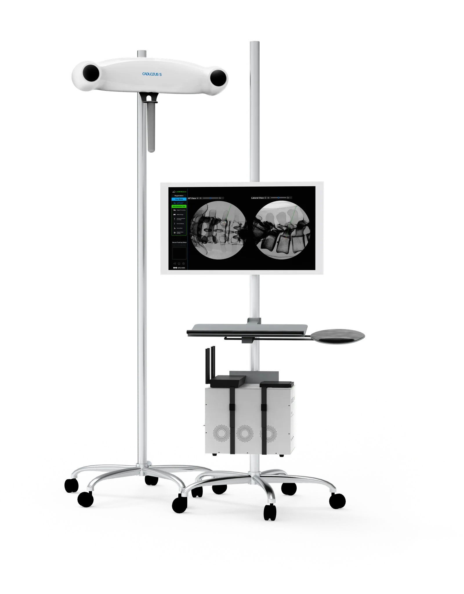
* Mean positional error: 1.80 mm in C-arm mode and 1.91 mm in CT mode; cadaver study, FDA 510(k) K220554.
AR navigation in action
Why augmented reality for spinal surgery?
Augmented reality merges the imaging with the real surgical environment rather than putting it on a screen beside it. That gives the surgeon real-time, intuitive guidance and a better-informed decision at every step. Six reasons it is where spinal navigation is going.
A live case, seen the way the surgeon sees it
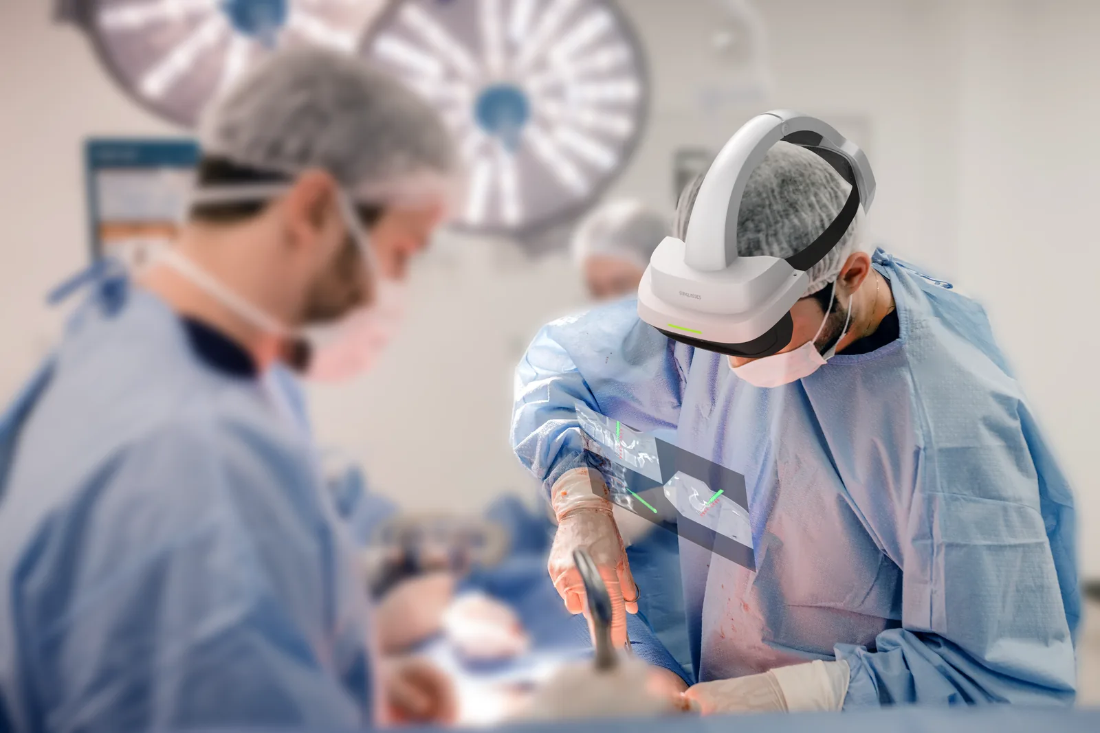
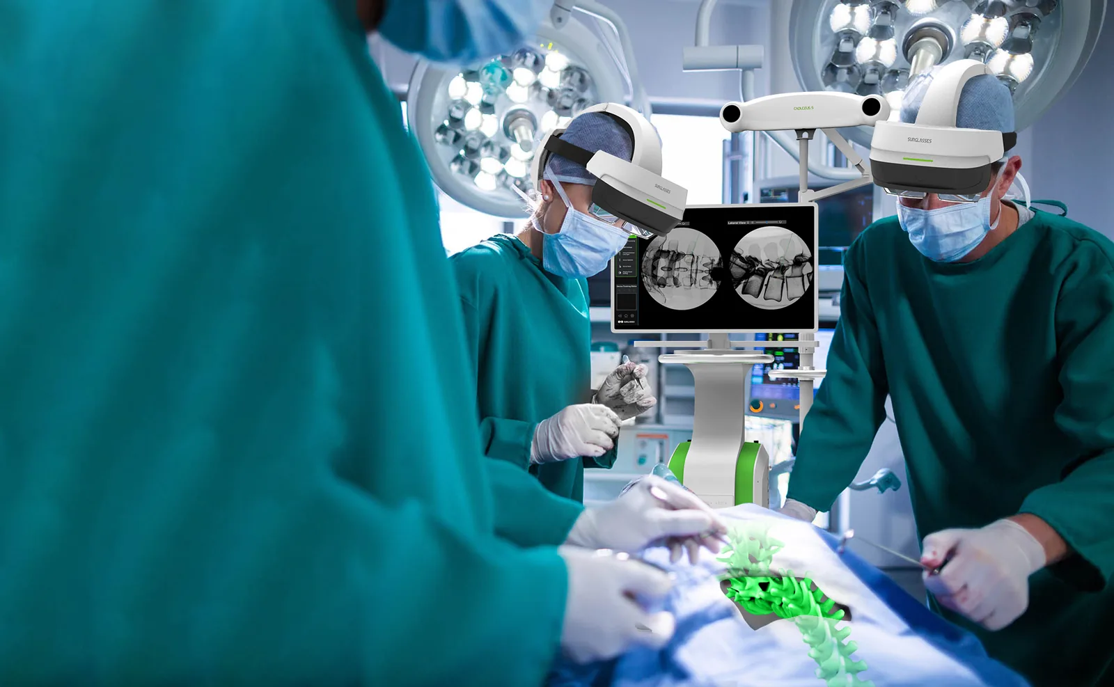
The spine model is registered onto the patient and stays there as the surgeon moves. Nothing on this footage is a mock-up — it is a real procedure in a real operating room.
The glance that AR removes
Conventional navigation asks the surgeon to look away from the wound to a screen on the far side of the room. Every glance costs focus. With Caduceus S the image is already in front of them.
Set up in 10 to 15 minutes
Registration is done from the C-arm images themselves, so the room is ready in 10 to 15 minutes rather than the thirty to forty a conventional system needs.
Calculated, not estimated
Critical imaging is projected into the surgeon's field of view, so instrument navigation is calculated rather than estimated. Mean positional error is under 2.0 mm (cadaver study, FDA 510(k) K220554).
Real-time guidance
Traditional navigation breaks the surgeon's concentration every time they turn to a monitor. Putting the data in their line of sight keeps them on the operative field and takes one class of human error out of the procedure.
Minimally invasive procedures
A clear view of the surgical area supports minimally invasive technique, through incisions of about 2 to 3 cm per segment.
Quick setup
Setup is streamlined to 10 to 15 minutes, against thirty to forty for conventional systems.
Four headsets at once
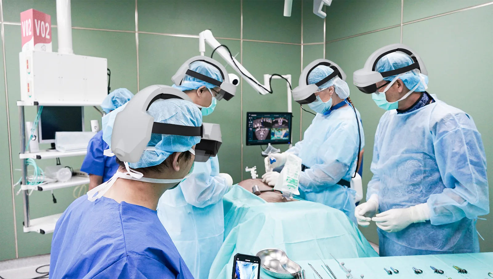
Up to four people — surgeon, assistants, scrub nurse, a rep — can wear the glasses simultaneously and see the same navigation. The case teaches the team while it is being done.
Works with what you already own
Caduceus S is built around existing hospital imaging, whether that is a 2D C-arm or 3D intraoperative imaging. Moving to AR-assisted surgery does not mean buying a new imaging suite first.
As AR matures its effect on spinal surgery grows rather than settles — better results, and a more efficient procedure for surgeon and patient alike. What that means clinically
Clinical benefits
What it changes, case after case
Precision is the headline, but the reasons surgeons keep the system in the room are more ordinary than that: the image is where they are looking, the whole team can see it, the instruments are the ones they already use, and the patient leaves with far less radiation than a conventional workflow would have given them.
Intuitive imaging, in the surgeon's own line of sight
The navigation image is displayed directly in the surgeon's field of view, which removes the need to look away at an external monitor to check position. Focus stays on the surgical site through the parts of the case where focus matters most.
The image itself arrives over a high-resolution wireless transmission system, so there is no cable run between the cart and the headset and no perceptible lag between what the instrument is doing and what the surgeon sees.
Up to four headsets in the same room
The system supports four headsets at once, so the surgeon, the assisting staff and anyone else scrubbed in are looking at the same navigation rather than at each other's description of it. Position can be discussed and confirmed out loud, in real time, by people who can all see the thing being discussed.
It also makes the case a teaching case at no extra cost — residents and nurses follow the anatomy as the surgeon works through it, instead of watching a monitor that is angled for somebody else.
Low radiation mode
Radiation dose is not a detail. It accumulates in the patient, and over a career it accumulates in the surgical team, raising the long-term risk of radiation-induced harm — including to bone marrow and the haematopoietic function that depends on it.
Caduceus S runs a low radiation mode. For patients and for everyone standing in the room, that is a large reduction in exposure for the same operation.
- No preoperative CT — the highest-dose scan in a conventional navigation workflow is not part of this one
- Two to four X-ray images taken before the incision to register the anatomy — and that is the imaging for the case
- No repeated confirmation shots during the procedure, which is where a conventional case quietly accumulates most of its dose
- The team benefits too — dose the patient does not receive is dose the surgeon and the scrub team do not stand in either
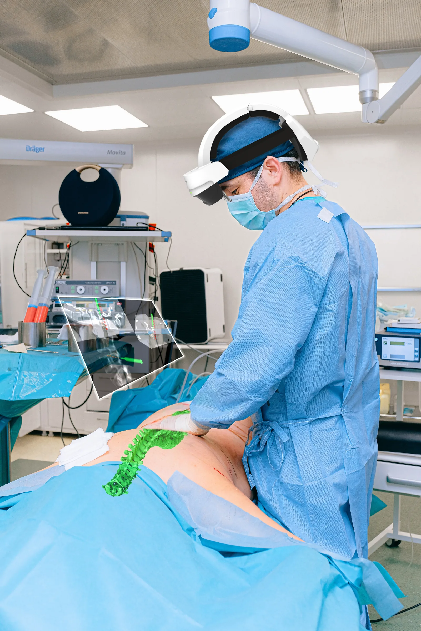
High-resolution wireless imaging
An advanced wireless transmission system delivers high-resolution images to the headset in real time, which is what makes navigation accurate rather than approximately accurate.
Built for long cases
Navigation holds up through a long case, so a difficult multi-level construct does not become a fight with the equipment.
Universal instruments
Caduceus S navigates with universal surgical instruments. No brand is mandated and nothing has to be swapped out, so the tray stays the one the surgeon knows.
Caduceus S is cleared by the US FDA under 510(k) and licensed as a Class II medical device by Taiwan's TFDA and the Thai FDA, and is manufactured under an ISO 13485:2016 quality management system. See every certification in full
Minimally invasive
Real MIS — 2 to 3 cm per segment, with the plan in view
Caduceus S is designed to raise the precision and the efficiency of minimally invasive spinal procedures. Whether the room has a 2D C-arm or 3D imaging, the system puts real-time, highly accurate information into the surgeon's line of sight rather than on a screen behind them.
Mean positional error under 2.0 mm
A mean positional error under 2.0 mm — 1.80 mm in C-arm mode and 1.91 mm in CT mode, in a cadaver study (FDA 510(k) K220554) — is the figure the rest of the system is built around.
- Distance, direction and angle to the target lesion, read live rather than inferred from two static views
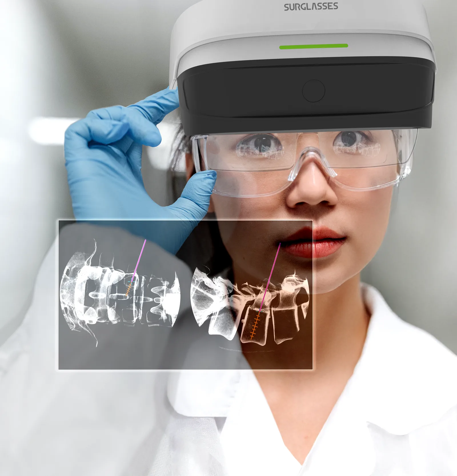
- Depth, on the way in — the trajectory and the graduated depth scale sit on the fluoroscopy image, so how far the instrument has travelled is a reading, not a judgement
- 2 to 3 cm per segment against 5 to 10 cm for traditional open spine surgery
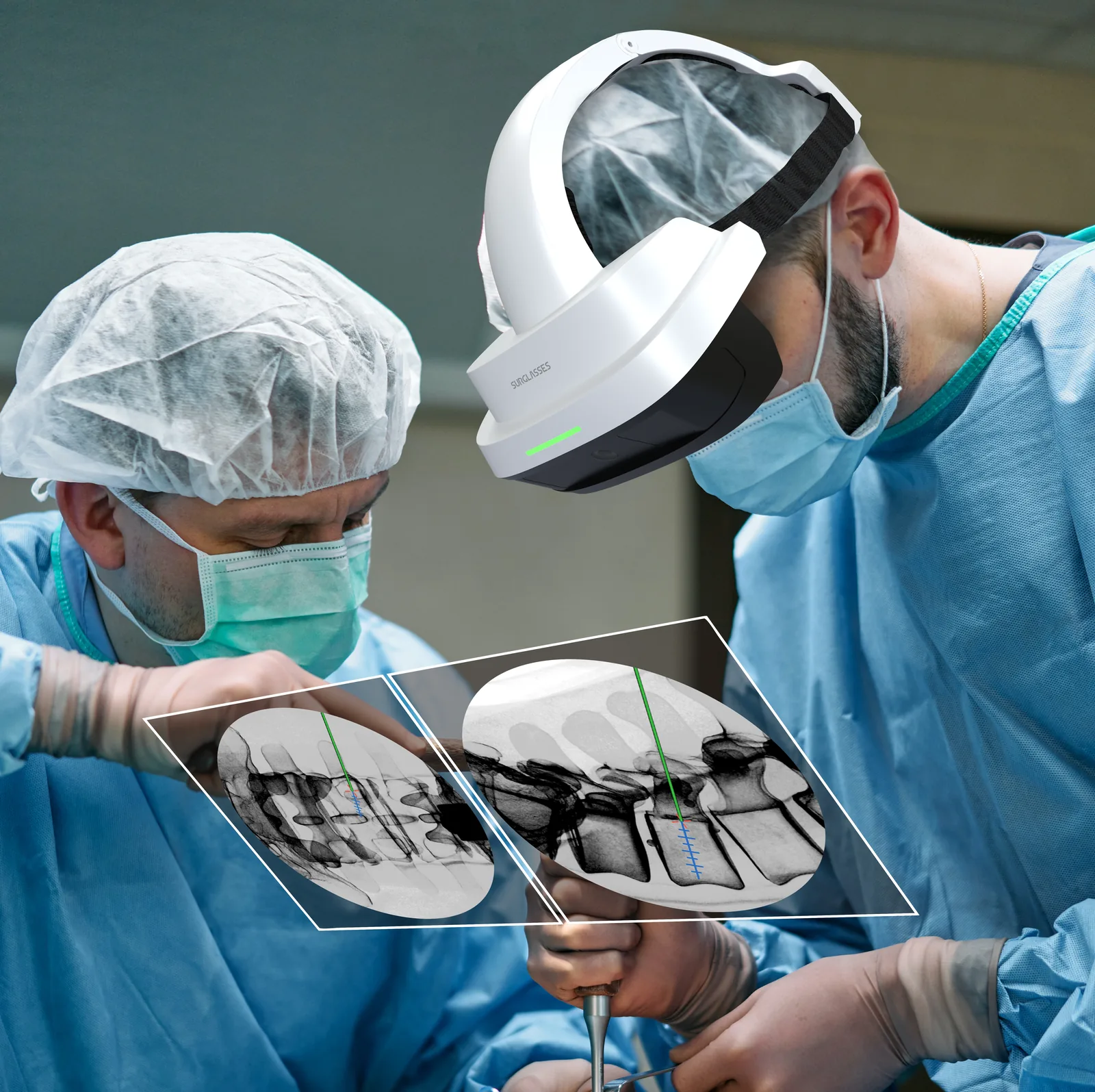
Seamless with 2D and 3D imaging — and switchable mid-case
The same headset renders a flat fluoroscopy panel, a 3D model registered onto the patient, or both together. Which one is in front of the surgeon is a choice made during the procedure, not a configuration decided when the system was bought.
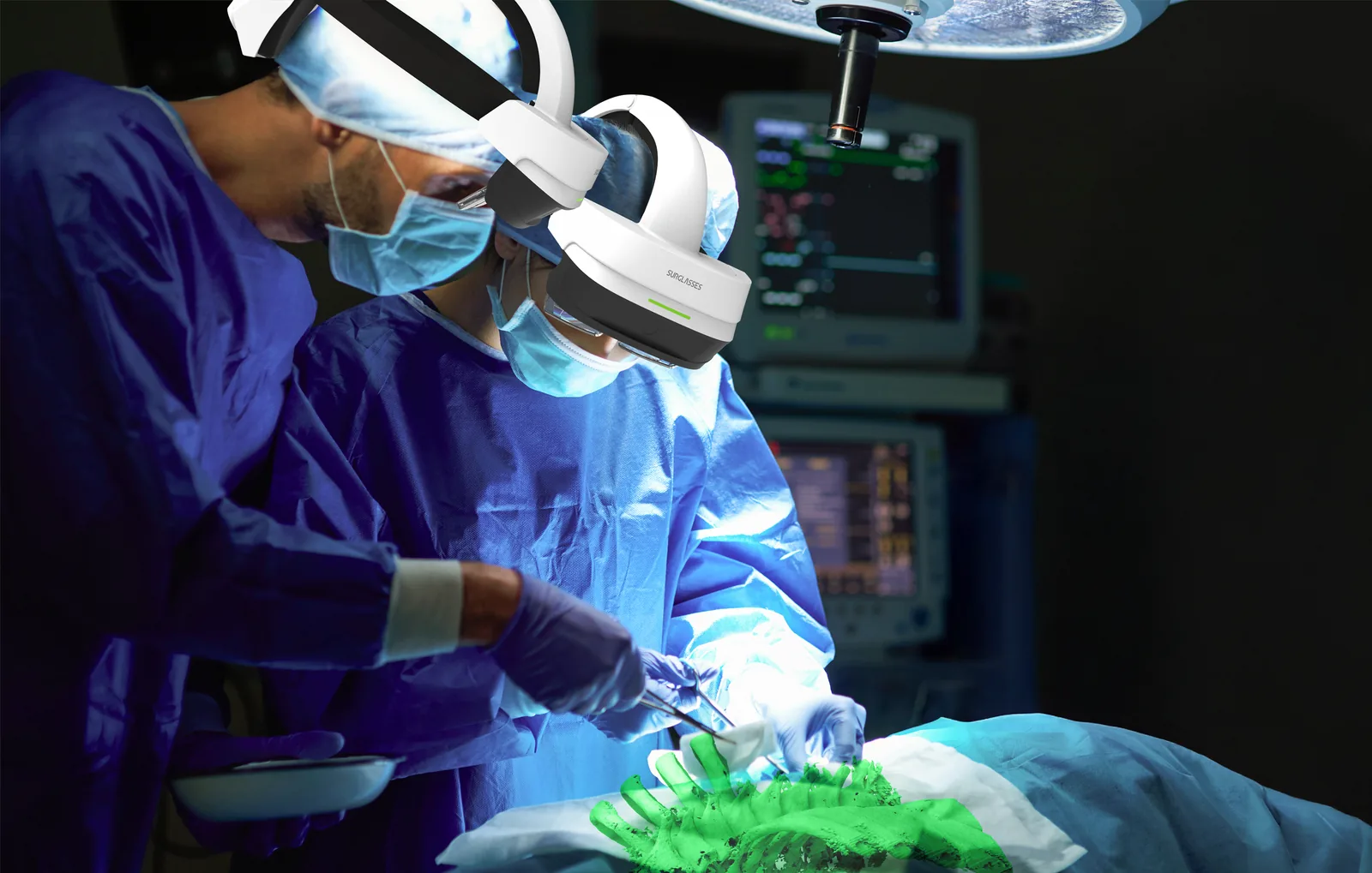
The plan, on the patient
A 3D model of the spine registered to the body itself, so the anatomy under the drape is visible in place rather than in the abstract.
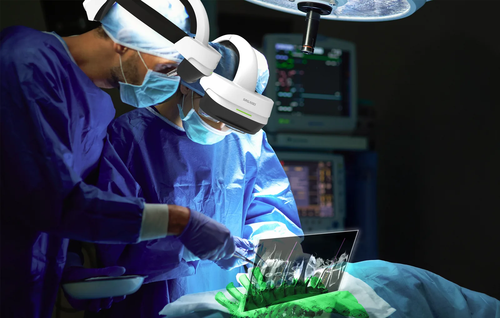
Both at once, when the step needs it
The registered model and the fluoroscopy panel together — the overview and the measurement in one field of view, with no screen equipment added to the room.
Fewer incisions to close, less blood lost and a faster return to normal life: the point of minimally invasive surgery has never been the size of the scar, it has been what the smaller scar makes possible.
Side by side
How it compares
Set against the two things a spine surgeon is choosing between — another navigation system, or open surgery without navigation at all — the differences are not marginal. They are in setup time, in radiation, in how long the patient stays, and in whether the surgeon has to look away from the wound.
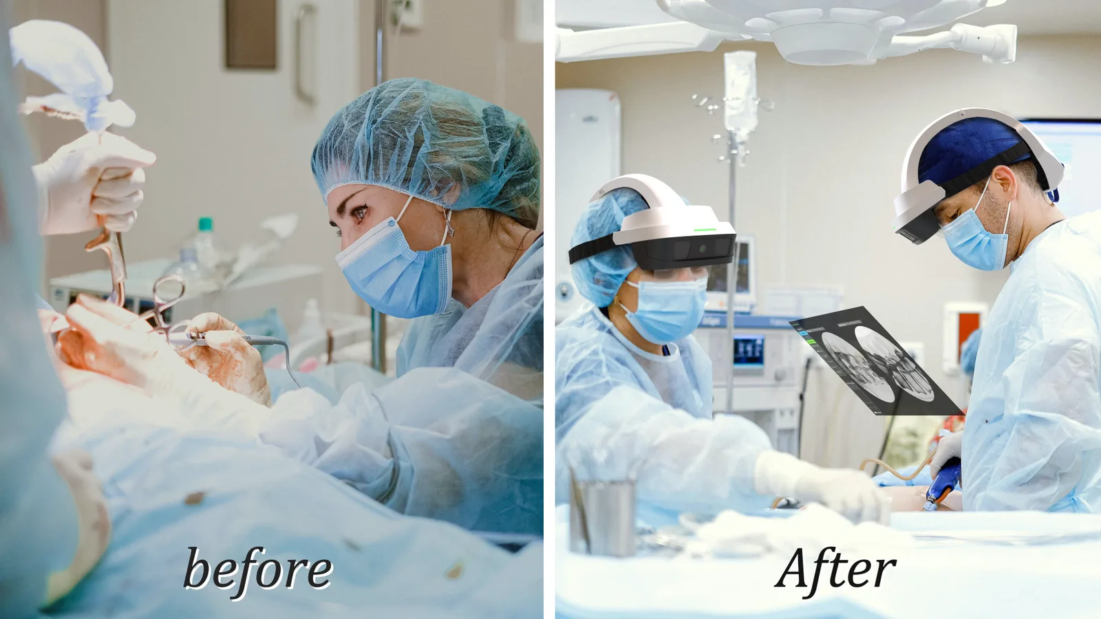
Before, and after
The change is easy to state and hard to overstate. On one side, a surgeon leaning into an open wound and turning to a monitor to check where they are. On the other, the same procedure with the imaging held in front of them and an incision small enough that most of it has stopped being an incision.
The figures in the table below are the ones the company measures its own system against. They are typical values for a single-segment case, not guarantees — every patient and every construct differs.
| Aspect | Caduceus S AR navigation | Other spine navigation systems | Traditional open spine surgery |
|---|---|---|---|
| Screen glances | None | Frequent | Frequent |
| Setup time | ~10–15 minutes | ~30–40 minutes | Not applicable |
| X-ray images required | 2–4 | Extensive | Extensive |
| Preoperative CT scan | Not required | Required | Not required |
| Incision size per segment | ~2–3 cm | ~2–3 cm | ~5–10 cm |
Typical values for a single-segment procedure, as measured by Surglasses. Actual results depend on the patient, the pathology and the construct.
Deployment settings
A hospital and an outpatient centre need different things
Caduceus S is one system with more than one shape. A hospital with 3D imaging or an intraoperative CT can navigate from a volume; an outpatient surgery centre with a single 2D C-arm can run the same AR-guided procedure without buying one.
Hospitals
For hospitals the system adds a 3D C-arm module and an intraoperative CT module — O-arm or a hybrid OR. It builds the 3D image during the case and projects it onto the surgical area through AR, and the surgeon can pick between three perspectives as the procedure demands.
Both modules attach to imaging equipment the hospital already owns, so the move to AR navigation is a change of workflow rather than a capital purchase. Which module runs is the hospital's choice: 2D C-arm for speed, intraoperative 3D for the cases that warrant it.
The individual navigation modules — 2D C-arm, 3D C-arm and Intra-CT — are described in detail on the Caduceus S navigation page.
Outpatient and ambulatory surgery centres
The case for OSCs and ASCs is largely financial. Caduceus S navigates from a standard 2D C-arm, so a centre can offer AR-guided spinal surgery without investing in 3D imaging. Two to four X-ray images before the incision, no intraoperative CT, and no second imaging device to find room for.
It adapts to the room rather than the other way round — large theatre or small procedure room — and the same system covers both traditional and minimally invasive navigation, so the choice stays with the surgeon and the patient in front of them.
Simpler workflow, lower cost per case, and precision that does not depend on equipment an outpatient centre would struggle to justify.
In either setting the clinical work is the same: pedicle screw insertion, scoliosis correction and vertebral fracture repair are what the system was built to navigate, and complex constructs are where AR earns its place.

Caduceus S — laptop version
Portable AR navigation
The same navigation, running on a laptop. Deployable across operating rooms, designed for speed, and light enough that moving it between theatres is not a decision anybody has to weigh up first.
One module, no cart
The laptop version supports one navigation module per software installation, chosen at purchase: the C-arm module, the 3D C-arm module, or the I-CT module as an option. Everything else — the AR projection, the sub-2.0 mm mean positional error, the two to four fluoroscopy images, the 2 to 3 cm incisions — is what the full system does.
- Quick bootstrapping — 10 to 15 minutes to set up, with minimal change to the existing workflow
- Compact and mobile — no bulky equipment, and no repeated table or C-arm height adjustments to get a usable image
- Fewer C-arm reshoots — the flow through the case is smoother, which is most of what surgeons notice first
- Spine implant and MIS procedures — the same two families of case the cart-based system is used for
Augmented reality, actual results
See it in your own operating room
Tell us what imaging you already have — a 2D C-arm, a 3D C-arm, an intraoperative CT — and the procedures you run most often, and we will show you the configuration that fits. Online, or on site with the system in front of you. If you are near our US office in Naples, Florida, we can bring the demo to you.