
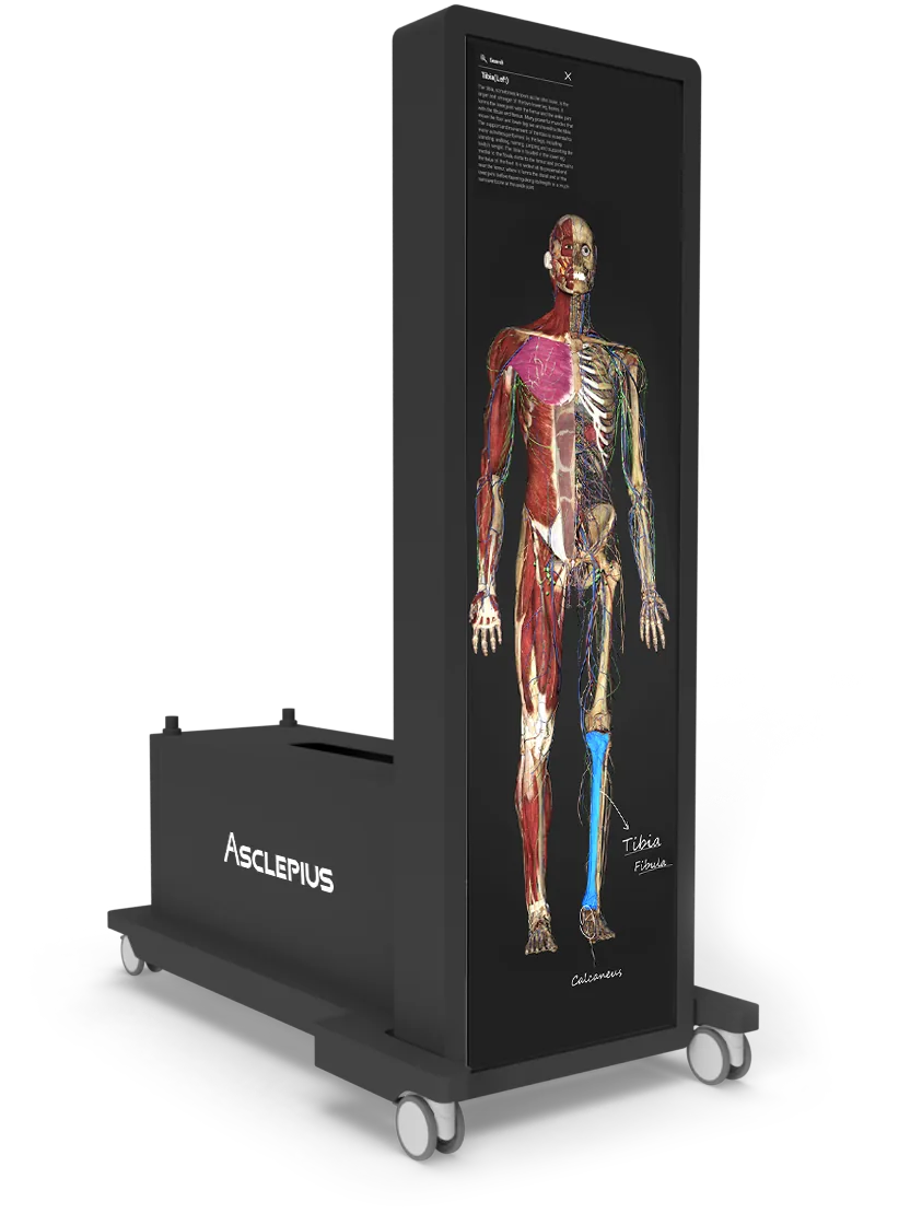
Asclepius Virtual Dissection Table
Dissect the same body as many times as the class needs
Asclepius 3.0 is a virtual dissection table built for medical education. Over 5,000 anatomical structures, 912 prepared teaching dissection views and a full clinical imaging library, on a touchscreen a whole group can gather around — in four sizes, from a mobile lecture table to a 99-inch teaching platform.
Asclepius 3.0
True anatomy, reconstructed structure by structure
Asclepius rebuilds the human body in 3D one anatomical structure at a time, so what appears on screen holds up to the same scrutiny as the specimen it was built from. Students interact with realistic digital anatomy instead of a cadaver — and they can start the dissection over as often as they like.
- Unlimited dissections — nothing is consumed, so a difficult region can be worked through again and again
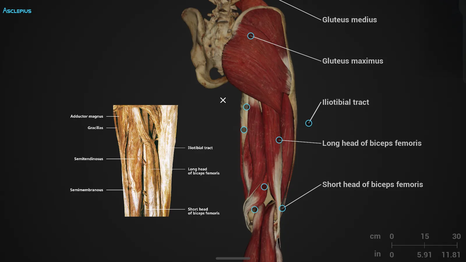
- 75 real-body comparison regions — the digital model held directly against a photograph of the real structure
- No formalin, no odour, no disposal — and none of the maintenance cost that comes with a wet lab
- Built with the people who teach from it — developed over years with clinicians and professors, and still expanding
What comes loaded
A teaching library, not just a model
Every Asclepius table ships with the whole content library — anatomy, imaging, slides, embryology, comparative anatomy, surgical planning and an examination bank, all kept up to date for the life of the table.
5,000+ anatomical structures
A digital human body with more than 5,000 individually modelled structures, each one separable from the layers around it.
912 teaching dissection views
Prepared 3D dissection perspectives, so the same region can be taught from whichever angle the point needs.
75 real-body comparison regions
High-precision comparison maps that place the real anatomical structure and its digital counterpart side by side.
1,000+ histology & pathology slides
High-resolution digital slides for studying tissue and pathology down to the cellular level.
Embryology, week 5 to 42
The complete anatomical growth sequence of the embryo and fetus, plus over 100 embryonic pathologies explained.
40 animal anatomy models
Forty species dissected in the same way as the human models, for comparative anatomy teaching.
DICOM imaging & 1,200+ clinical studies
Render CT and MRI data in DICOM format, or work from a database of real clinical orthopaedic studies.
Surgical simulation & planning
Educational preoperative planning with customisable bone plates and screws, spinal puncture and dental implant simulation.
Tap a structure, get its name
Every anatomical part can be clicked for its definition and explanation, at the resolution needed to read the smallest detail.
Thousands of resources, one menu
Over a thousand pieces of anatomical and physiological reference material sit behind the same panel, system by system.
Growth stages you can rotate
Fetal development from week 5 to week 42, rendered in 3D rather than shown as a sequence of textbook plates.
Rich and unique teaching content
Six software packages on one table
Four packages are included with every table from 65 inches up, and two more can be added. Each carries its own anatomical figures, imaging and surgical cases, and each is updated throughout the warranty period at no extra cost.
Anatomy
The core human anatomy library — every structure separable, every layer removable, with teaching dissection views prepared for class use.
OOOPDS Radiology
DICOM CT and MRI studies read alongside the anatomical model, backed by a library of real clinical orthopaedic cases.

Histopathology Atlas
Over a thousand high-definition digital slides, zoomable to cellular level for tissue and pathology teaching.
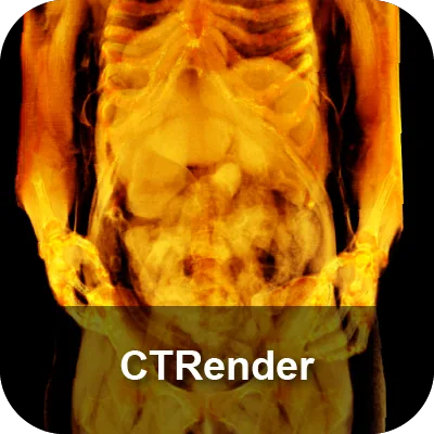
CTRender
Volume rendering of CT and MRI data, so a real patient study can be turned into a three-dimensional object the class can turn over.
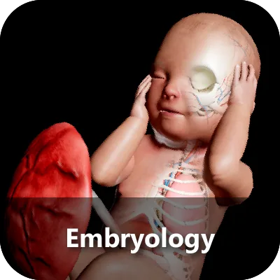
Embryology
Optional. Fetal development from week 5 to week 42, with more than 100 embryonic pathologies described.
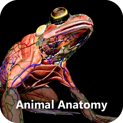
Animal Anatomy
Optional. Forty species for comparative anatomy, dissected to the same standard as the human models.
Technologies
What the table is doing underneath
A touchscreen, a rendering engine and a content platform, put together so that the person teaching never has to think about any of them.
Interactive touchscreen
An intuitive touch interface for engaging with and manipulating the anatomical models directly, with immediate control and no intermediate device.
High-resolution imaging
Ultra-high-definition rendering of anatomical structures, precise enough for clinical use as well as teaching, with a definition behind every click.
Thousands of resources
An extensive library of anatomical and physiological reference material, searchable from the same panel the models are driven from.
Real clinical cases
Built-in clinical case studies bridge theory and practice — and teaching staff can upload their own imaging to teach from.
Nursing & medical testing
Examination modules built for nursing and medical assessment, drawn from historical professional licensure papers.
Updates under warranty
New features and content arrive as software updates throughout the warranty period, at no additional cost.
Models
Choose the fit for your room, not for the brochure
Four sizes of the same platform, sized by how many people need to stand around the table at once. The 43-inch model moves between classrooms on its own castors; the 84 and 99-inch models rise from flat to upright on a single button, so a whole cohort can see a body at life scale.
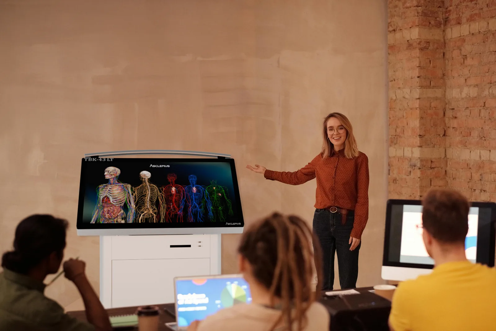
TBK-43 LT
Lecture table · 2–4 people. The most compact model in the series, and it carries a 4K panel. Small classrooms, compact laboratories and teacher-assisted podiums; light enough to be wheeled between rooms for mobile teaching.
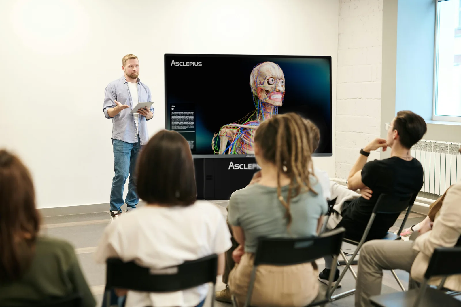
TBK-65 4K
Small group · 5–7 people. A 4K panel at a size a small group can gather round, which suits histology and pathology work — finer cell tissue stays readable. Tilts electronically to any angle.
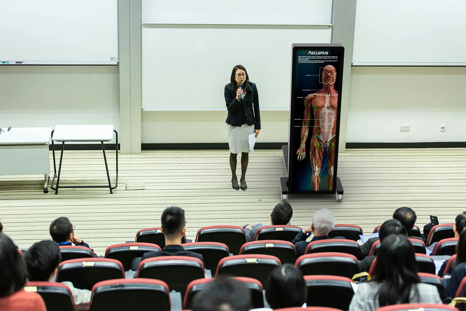
TBK-84 EA
Larger group · 7–10 people. Built for large classrooms and anatomy laboratories. Remote-controlled electronic lifting switches the screen between upright and flat with one button.
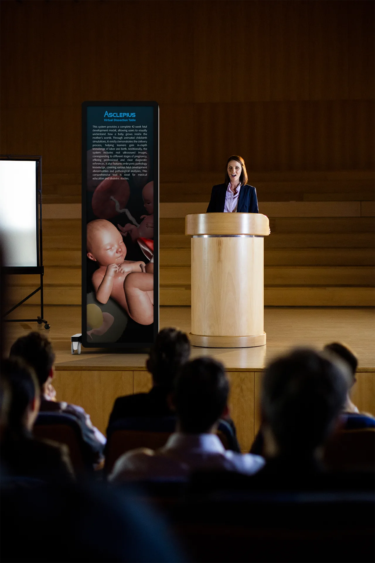
TBK-99 EA
Large-scale course · 7–10 people. The largest digital touch anatomy table. Anatomical figures are shown at life scale, so nobody in the room has to imagine the size of anything.
Specifications side by side
| Specification | TBK-43 LT | TBK-65 4K | TBK-84 EA | TBK-99 EA |
|---|---|---|---|---|
| Screen size | 43 inches | 65 inches | 84 inches | 99 inches |
| Tilt angle | 0° – 45° | 0° – 90° electronically adjustable |
0° & 90° electronically adjustable |
0° & 90° electronically adjustable |
| Group size | 2–4 people | 5–7 people | 7–10 people | 7–10 people |
| Software included | Anatomy, OOOPDS Radiology | Anatomy, OOOPDS Radiology, Histopathology Atlas, CTRender | Anatomy, OOOPDS Radiology, Histopathology Atlas, CTRender | Anatomy, OOOPDS Radiology, Histopathology Atlas, CTRender |
Embryology and Animal Anatomy are available as options on the 65, 84 and 99-inch models. The 43-inch lecture table ships with Anatomy and OOOPDS Radiology.
For the full specification of any model — panel resolution, dimensions flat and upright, and weight — tell us which model you are considering on the contact page and we will send you its specification sheet.
Looking for the AI version instead? See the Asclepius AI Table
See it for yourself
Put a body on the table and take it apart
Tell us the courses you run, the size of your groups and the room you have, and we will show you which of the four models fits and which software packages you actually need — online, or on site with the table in front of you.